 Movie
Movie Controller

[English] 日本語
 Yorodumi
- PDB-6rx1: Crystal structure of human syncytin 1 in post-fusion conformation -
+ Open data

- Basic information
| Entry | Database: PDB / ID: 6rx1 | ||||||
|---|---|---|---|---|---|---|---|
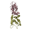
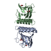
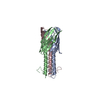
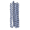
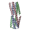
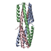
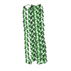
| Title | Crystal structure of human syncytin 1 in post-fusion conformation | ||||||
 Components Components | Syncytin-1 | ||||||
 Keywords Keywords | MEMBRANE PROTEIN / HUMAN PLACENTAL PROTEIN / MEMBRANE FUSION / ENDOGENOUS RETROVIRUS / HERV-W / SYNCYTIN | ||||||
| Function / homology |  Function and homology information Function and homology informationsyncytium formation by cell-cell fusion / : / myoblast fusion / anatomical structure morphogenesis / plasma membrane Similarity search - Function | ||||||
| Biological species |  Homo sapiens (human) Homo sapiens (human) | ||||||
| Method |  X-RAY DIFFRACTION / SYNCHROTRON / MOLECULAR REPLACEMENT / Resolution: 2.1 Å X-RAY DIFFRACTION / SYNCHROTRON / MOLECULAR REPLACEMENT / Resolution: 2.1 Å | ||||||
 Authors Authors | Ruigrok, K. / Backovic, M. / Vaney, M.C. / Rey, F.A. | ||||||
| Funding support |  France, 1items France, 1items
| ||||||
 Citation Citation | Journal: J.Mol.Biol. / Year: 2019 Title: X-ray Structures of the Post-fusion 6-Helix Bundle of the Human Syncytins and their Functional Implications. Authors: Ruigrok, K. / Vaney, M.C. / Buchrieser, J. / Baquero, E. / Hellert, J. / Baron, B. / England, P. / Schwartz, O. / Rey, F.A. / Backovic, M. #1: Journal: J.Mol.Biol. / Year: 2005Title: Crystal structure of a pivotal domain of human syncytin-2, a 40 million years old endogenous retrovirus fusogenic envelope gene captured by primates. Authors: Renard, M. / Varela, P.F. / Letzelter, C. / Duquerroy, S. / Rey, F.A. / Heidmann, T. | ||||||
| History |
|
- Structure visualization
| Structure viewer | Molecule: MolmilJmol/JSmol |
|---|
- Downloads & links
-Download
| PDBx/mmCIF format | 6rx1.cif.gz | 35.1 KB | Display | PDBx/mmCIF format |
|---|---|---|---|---|
| PDB format | pdb6rx1.ent.gz | 21.9 KB | Display | PDB format |
| PDBx/mmJSON format | 6rx1.json.gz | Tree view | PDBx/mmJSON format | |
| Others |  Other downloads Other downloads |
-Validation report
| Arichive directory | https://data.pdbj.org/pub/pdb/validation_reports/rx/6rx1ftp://data.pdbj.org/pub/pdb/validation_reports/rx/6rx1 | HTTPS FTP |
|---|
-Related structure data
| Related structure data |  6rx3C  1y4mS S: Starting model for refinement C: citing same article ( |
|---|---|
| Similar structure data |


-Links
 PDBj
PDBj- Assembly
| Deposited unit | 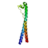
| |||||||||
|---|---|---|---|---|---|---|---|---|---|---|
| 1 | 
| |||||||||
| Unit cell |
| |||||||||
| Components on special symmetry positions |
|
-Components
| #1: Protein | Mass: 12614.133 Da / Num. of mol.: 1 Source method: isolated from a genetically manipulated source Details: The following sequence MHHHHHHENLYFQS at the N-terminal of the protein sequence is from an expression tag with the 1st residue MET as the initiating methionine. The 3 first residues 'TST' ...Details: The following sequence MHHHHHHENLYFQS at the N-terminal of the protein sequence is from an expression tag with the 1st residue MET as the initiating methionine. The 3 first residues 'TST' from the protein sequence are disordered in density. Source: (gene. exp.) Homo sapiens (human) / Gene: ERVW-1, ERVWE1 / Plasmid: PET28a / Production host:  |
|---|---|
| #2: Chemical | ChemComp-GOL /   Mass: 92.094 Da / Num. of mol.: 1 / Source method: obtained synthetically / Formula: C3H8O3 / Feature type: SUBJECT OF INVESTIGATION Mass: 92.094 Da / Num. of mol.: 1 / Source method: obtained synthetically / Formula: C3H8O3 / Feature type: SUBJECT OF INVESTIGATION |
| #3: Chemical | ChemComp-CL /   Mass: 35.453 Da / Num. of mol.: 1 / Source method: isolated from a natural source / Formula: Cl Mass: 35.453 Da / Num. of mol.: 1 / Source method: isolated from a natural source / Formula: Cl |
| #4: Water | ChemComp-HOH /  Mass: 18.015 Da / Num. of mol.: 70 / Source method: isolated from a natural source / Formula: H2O Mass: 18.015 Da / Num. of mol.: 70 / Source method: isolated from a natural source / Formula: H2O |
| Has ligand of interest | N |
| Has protein modification | Y |
-Experimental details
-Experiment
| Experiment | Method: X-RAY DIFFRACTION / Number of used crystals: 1 |
|---|
- Sample preparation
| Crystal | Density Matthews: 2.52 Å3/Da / Density % sol: 51.16 % |
|---|---|
| Crystal grow | Temperature: 295 K / Method: vapor diffusion, sitting drop / pH: 7.5 Details: 0.1 M HEPES pH 7.5, 30% v/v 2-propanol, 0.2 M MgCl2 |
-Data collection
| Diffraction | Mean temperature: 100 K / Serial crystal experiment: N |
|---|---|
| Diffraction source | Source: SYNCHROTRON / Site: SOLEIL / Beamline: PROXIMA 2 / Wavelength: 1.070638 Å |
| Detector | Type: DECTRIS EIGER X 9M / Detector: PIXEL / Date: Dec 17, 2016 |
| Radiation | Monochromator: cryogenically cooled channel cut crystal monochromator, a convex prefocussing mirror and a KirkpatrickBaez pair of focussing mirrors Protocol: SINGLE WAVELENGTH / Monochromatic (M) / Laue (L): M / Scattering type: x-ray |
| Radiation wavelength | Wavelength: 1.070638 Å / Relative weight: 1 |
| Reflection | Resolution: 2.1→43.1 Å / Num. obs: 7752 / % possible obs: 99.1 % / Redundancy: 5.8 % / Biso Wilson estimate: 30 Å2 / CC1/2: 0.99 / Rmerge(I) obs: 0.12 / Rpim(I) all: 0.08 / Rrim(I) all: 0.15 / Net I/σ(I): 7 |
| Reflection shell | Resolution: 2.1→2.16 Å / Redundancy: 6.1 % / Rmerge(I) obs: 1.25 / Mean I/σ(I) obs: 1.1 / Num. unique obs: 622 / CC1/2: 0.47 / Rpim(I) all: 0.81 / Rrim(I) all: 1.5 / % possible all: 99.9 |
- Processing
| Software |
| ||||||||||||||||||||||||||||||||||||||||||||||||||||||||||||||||||||||||||||||||||||||||||||||||||||||||||||
|---|---|---|---|---|---|---|---|---|---|---|---|---|---|---|---|---|---|---|---|---|---|---|---|---|---|---|---|---|---|---|---|---|---|---|---|---|---|---|---|---|---|---|---|---|---|---|---|---|---|---|---|---|---|---|---|---|---|---|---|---|---|---|---|---|---|---|---|---|---|---|---|---|---|---|---|---|---|---|---|---|---|---|---|---|---|---|---|---|---|---|---|---|---|---|---|---|---|---|---|---|---|---|---|---|---|---|---|---|---|
| Refinement | Method to determine structure: MOLECULAR REPLACEMENT Starting model: 1Y4M Resolution: 2.1→20 Å / Cor.coef. Fo:Fc: 0.941 / Cor.coef. Fo:Fc free: 0.935 / SU R Cruickshank DPI: 0.19 / Cross valid method: THROUGHOUT / σ(F): 0 / SU R Blow DPI: 0.212 / SU Rfree Blow DPI: 0.172 / SU Rfree Cruickshank DPI: 0.164
| ||||||||||||||||||||||||||||||||||||||||||||||||||||||||||||||||||||||||||||||||||||||||||||||||||||||||||||
| Displacement parameters | Biso max: 154.08 Å2 / Biso mean: 50.38 Å2 / Biso min: 30.2 Å2
| ||||||||||||||||||||||||||||||||||||||||||||||||||||||||||||||||||||||||||||||||||||||||||||||||||||||||||||
| Refine analyze | Luzzati coordinate error obs: 0.33 Å | ||||||||||||||||||||||||||||||||||||||||||||||||||||||||||||||||||||||||||||||||||||||||||||||||||||||||||||
| Refinement step | Cycle: final / Resolution: 2.1→20 Å
| ||||||||||||||||||||||||||||||||||||||||||||||||||||||||||||||||||||||||||||||||||||||||||||||||||||||||||||
| Refine LS restraints |
| ||||||||||||||||||||||||||||||||||||||||||||||||||||||||||||||||||||||||||||||||||||||||||||||||||||||||||||
| LS refinement shell | Resolution: 2.1→2.35 Å / Rfactor Rfree error: 0 / Total num. of bins used: 5
|
