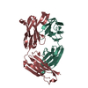
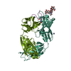
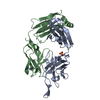
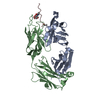
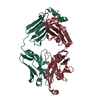
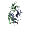
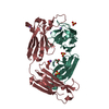
ジャーナル: PLoS Pathog / 年: 2013 タイトル: Broadly neutralizing antibody PGT121 allosterically modulates CD4 binding via recognition of the HIV-1 gp120 V3 base and multiple surrounding glycans. 著者: Jean-Philippe Julien / Devin Sok / Reza Khayat / Jeong Hyun Lee / Katie J Doores / Laura M Walker / Alejandra Ramos / Devan C Diwanji / Robert Pejchal / Albert Cupo / Umesh Katpally / Rafael ...著者: Jean-Philippe Julien / Devin Sok / Reza Khayat / Jeong Hyun Lee / Katie J Doores / Laura M Walker / Alejandra Ramos / Devan C Diwanji / Robert Pejchal / Albert Cupo / Umesh Katpally / Rafael S Depetris / Robyn L Stanfield / Ryan McBride / Andre J Marozsan / James C Paulson / Rogier W Sanders / John P Moore / Dennis R Burton / Pascal Poignard / Andrew B Ward / Ian A Wilson / 要旨: New broad and potent neutralizing HIV-1 antibodies have recently been described that are largely dependent on the gp120 N332 glycan for Env recognition. Members of the PGT121 family of antibodies, ...New broad and potent neutralizing HIV-1 antibodies have recently been described that are largely dependent on the gp120 N332 glycan for Env recognition. Members of the PGT121 family of antibodies, isolated from an African donor, neutralize ∼70% of circulating isolates with a median IC50 less than 0.05 µg ml(-1). Here, we show that three family members, PGT121, PGT122 and PGT123, have very similar crystal structures. A long 24-residue HCDR3 divides the antibody binding site into two functional surfaces, consisting of an open face, formed by the heavy chain CDRs, and an elongated face, formed by LCDR1, LCDR3 and the tip of the HCDR3. Alanine scanning mutagenesis of the antibody paratope reveals a crucial role in neutralization for residues on the elongated face, whereas the open face, which accommodates a complex biantennary glycan in the PGT121 structure, appears to play a more secondary role. Negative-stain EM reconstructions of an engineered recombinant Env gp140 trimer (SOSIP.664) reveal that PGT122 interacts with the gp120 outer domain at a more vertical angle with respect to the top surface of the spike than the previously characterized antibody PGT128, which is also dependent on the N332 glycan. We then used ITC and FACS to demonstrate that the PGT121 antibodies inhibit CD4 binding to gp120 despite the epitope being distal from the CD4 binding site. Together, these structural, functional and biophysical results suggest that the PGT121 antibodies may interfere with Env receptor engagement by an allosteric mechanism in which key structural elements, such as the V3 base, the N332 oligomannose glycan and surrounding glycans, including a putative V1/V2 complex biantennary glycan, are conformationally constrained.
モノクロメーター: Side scattering bent cube-root I-beam single crystal, asymmetric cut 4.965 degrees プロトコル: SINGLE WAVELENGTH / 単色(M)・ラウエ(L): M / 散乱光タイプ: x-ray
 ムービー
ムービー コントローラー


 データを開く
 基本情報
基本情報 要素
要素 キーワード
キーワード 機能・相同性情報
 Homo sapiens (ヒト)
 X線回折 /
  データ登録者
 引用
引用
 構造の表示
構造の表示 ダウンロードとリンク
 その他のダウンロード


 PDBj
PDBj

 集合体
集合体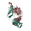

 分子量: 92.094 Da / 分子数: 3 / 由来タイプ: 合成 / 式: C3H8O3
 分子量: 18.015 Da / 分子数: 248 / 由来タイプ: 天然 / 式: H2O
 試料調製
試料調製 解析
解析