 Movie
Movie Controller

[English] 日本語
 Yorodumi
- PDB-4jy5: Crystal structure of human Fab PGT122, a broadly reactive and pot... -
+ Open data

- Basic information
| Entry | Database: PDB / ID: 4jy5 | ||||||
|---|---|---|---|---|---|---|---|
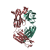
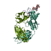
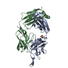
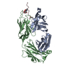
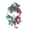
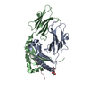
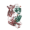
| Title | Crystal structure of human Fab PGT122, a broadly reactive and potent HIV-1 neutralizing antibody | ||||||
 Components Components |
| ||||||
 Keywords Keywords | IMMUNE SYSTEM / Broadly neutralizing antibody against HIV-1 / HIV-1 Env gp120 subunit | ||||||
| Function / homology | Immunoglobulins / Immunoglobulin-like / Sandwich / Mainly Beta Function and homology information Function and homology information | ||||||
| Biological species |  Homo sapiens (human) Homo sapiens (human) | ||||||
| Method |  X-RAY DIFFRACTION / SYNCHROTRON / MOLECULAR REPLACEMENT / Resolution: 1.75 Å X-RAY DIFFRACTION / SYNCHROTRON / MOLECULAR REPLACEMENT / Resolution: 1.75 Å | ||||||
 Authors Authors | Julien, J.-P. / Diwanji, D.C. / Burton, D.R. / Wilson, I.A. | ||||||
 Citation Citation | Journal: PLoS Pathog / Year: 2013 Title: Broadly neutralizing antibody PGT121 allosterically modulates CD4 binding via recognition of the HIV-1 gp120 V3 base and multiple surrounding glycans. Authors: Jean-Philippe Julien / Devin Sok / Reza Khayat / Jeong Hyun Lee / Katie J Doores / Laura M Walker / Alejandra Ramos / Devan C Diwanji / Robert Pejchal / Albert Cupo / Umesh Katpally / Rafael ...Authors: Jean-Philippe Julien / Devin Sok / Reza Khayat / Jeong Hyun Lee / Katie J Doores / Laura M Walker / Alejandra Ramos / Devan C Diwanji / Robert Pejchal / Albert Cupo / Umesh Katpally / Rafael S Depetris / Robyn L Stanfield / Ryan McBride / Andre J Marozsan / James C Paulson / Rogier W Sanders / John P Moore / Dennis R Burton / Pascal Poignard / Andrew B Ward / Ian A Wilson /  Abstract: New broad and potent neutralizing HIV-1 antibodies have recently been described that are largely dependent on the gp120 N332 glycan for Env recognition. Members of the PGT121 family of antibodies, ...New broad and potent neutralizing HIV-1 antibodies have recently been described that are largely dependent on the gp120 N332 glycan for Env recognition. Members of the PGT121 family of antibodies, isolated from an African donor, neutralize ∼70% of circulating isolates with a median IC50 less than 0.05 µg ml(-1). Here, we show that three family members, PGT121, PGT122 and PGT123, have very similar crystal structures. A long 24-residue HCDR3 divides the antibody binding site into two functional surfaces, consisting of an open face, formed by the heavy chain CDRs, and an elongated face, formed by LCDR1, LCDR3 and the tip of the HCDR3. Alanine scanning mutagenesis of the antibody paratope reveals a crucial role in neutralization for residues on the elongated face, whereas the open face, which accommodates a complex biantennary glycan in the PGT121 structure, appears to play a more secondary role. Negative-stain EM reconstructions of an engineered recombinant Env gp140 trimer (SOSIP.664) reveal that PGT122 interacts with the gp120 outer domain at a more vertical angle with respect to the top surface of the spike than the previously characterized antibody PGT128, which is also dependent on the N332 glycan. We then used ITC and FACS to demonstrate that the PGT121 antibodies inhibit CD4 binding to gp120 despite the epitope being distal from the CD4 binding site. Together, these structural, functional and biophysical results suggest that the PGT121 antibodies may interfere with Env receptor engagement by an allosteric mechanism in which key structural elements, such as the V3 base, the N332 oligomannose glycan and surrounding glycans, including a putative V1/V2 complex biantennary glycan, are conformationally constrained. | ||||||
| History |
|
- Structure visualization
| Structure viewer | Molecule: MolmilJmol/JSmol |
|---|
- Downloads & links
-Download
| PDBx/mmCIF format | 4jy5.cif.gz | 186.9 KB | Display | PDBx/mmCIF format |
|---|---|---|---|---|
| PDB format | pdb4jy5.ent.gz | 147.6 KB | Display | PDB format |
| PDBx/mmJSON format | 4jy5.json.gz | Tree view | PDBx/mmJSON format | |
| Others |  Other downloads Other downloads |
-Validation report
| Arichive directory | https://data.pdbj.org/pub/pdb/validation_reports/jy/4jy5ftp://data.pdbj.org/pub/pdb/validation_reports/jy/4jy5 | HTTPS FTP |
|---|
-Related structure data
| Related structure data |  5624C  4jy4C  4jy6SC C: citing same article ( S: Starting model for refinement |
|---|---|
| Similar structure data |


-Links
 PDBj
PDBj

- Assembly
| Deposited unit | 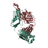
| ||||||||
|---|---|---|---|---|---|---|---|---|---|
| 1 |
| ||||||||
| Unit cell |
|
-Components
| #1: Antibody | Mass: 22712.082 Da / Num. of mol.: 1 / Fragment: Fab Source method: isolated from a genetically manipulated source Source: (gene. exp.) Homo sapiens (human) / Cell line (production host): HEK 293F / Production host: Homo sapiens (human) | ||||
|---|---|---|---|---|---|
| #2: Antibody | Mass: 25434.691 Da / Num. of mol.: 1 / Fragment: Fab Source method: isolated from a genetically manipulated source Source: (gene. exp.) Homo sapiens (human) / Cell line (production host): HEK 293F / Production host: Homo sapiens (human) | ||||
| #3: Chemical |   Mass: 92.094 Da / Num. of mol.: 3 / Source method: obtained synthetically / Formula: C3H8O3 Mass: 92.094 Da / Num. of mol.: 3 / Source method: obtained synthetically / Formula: C3H8O3#4: Water | ChemComp-HOH / |  Mass: 18.015 Da / Num. of mol.: 248 / Source method: isolated from a natural source / Formula: H2O Mass: 18.015 Da / Num. of mol.: 248 / Source method: isolated from a natural source / Formula: H2OHas protein modification | Y | |
-Experimental details
-Experiment
| Experiment | Method: X-RAY DIFFRACTION / Number of used crystals: 1 |
|---|
- Sample preparation
| Crystal | Density Matthews: 2.04 Å3/Da / Density % sol: 39.56 % |
|---|---|
| Crystal grow | Temperature: 293 K / Method: vapor diffusion, sitting drop / pH: 10.5 Details: 0.1 M CAPS, pH 10.5, 0.2 M sodium chloride, 20% w/v PEG8000, VAPOR DIFFUSION, SITTING DROP, temperature 293K |
-Data collection
| Diffraction | Mean temperature: 100 K |
|---|---|
| Diffraction source | Source: SYNCHROTRON / Site: SSRL / Beamline: BL11-1 / Wavelength: 0.979 Å |
| Detector | Type: PSI PILATUS 6M / Detector: PIXEL / Date: May 5, 2012 |
| Radiation | Monochromator: Side scattering bent cube-root I-beam single crystal, asymmetric cut 4.965 degrees Protocol: SINGLE WAVELENGTH / Monochromatic (M) / Laue (L): M / Scattering type: x-ray |
| Radiation wavelength | Wavelength: 0.979 Å / Relative weight: 1 |
| Reflection | Resolution: 1.75→40 Å / Num. all: 39317 / Num. obs: 38560 / % possible obs: 97.7 % / Observed criterion σ(F): 0 / Observed criterion σ(I): 0 / Redundancy: 3.3 % / Rsym value: 0.064 / Net I/σ(I): 11.4 |
| Reflection shell | Resolution: 1.75→1.85 Å / Redundancy: 3.2 % / Mean I/σ(I) obs: 2.4 / Num. unique all: 5963 / Rsym value: 0.43 / % possible all: 97.2 |
- Processing
| Software |
| |||||||||||||||||||||||||||||||||||||||||||||||||||||||||||||||||||||||||||||||||||||||||||||||||||||||||||||||||||||||||||||||||||||||||||||||||||||||||||||||||||||||||||||||||||||||||||||
|---|---|---|---|---|---|---|---|---|---|---|---|---|---|---|---|---|---|---|---|---|---|---|---|---|---|---|---|---|---|---|---|---|---|---|---|---|---|---|---|---|---|---|---|---|---|---|---|---|---|---|---|---|---|---|---|---|---|---|---|---|---|---|---|---|---|---|---|---|---|---|---|---|---|---|---|---|---|---|---|---|---|---|---|---|---|---|---|---|---|---|---|---|---|---|---|---|---|---|---|---|---|---|---|---|---|---|---|---|---|---|---|---|---|---|---|---|---|---|---|---|---|---|---|---|---|---|---|---|---|---|---|---|---|---|---|---|---|---|---|---|---|---|---|---|---|---|---|---|---|---|---|---|---|---|---|---|---|---|---|---|---|---|---|---|---|---|---|---|---|---|---|---|---|---|---|---|---|---|---|---|---|---|---|---|---|---|---|---|---|---|
| Refinement | Method to determine structure: MOLECULAR REPLACEMENT Starting model: PDB ENTRY 4JY6 Resolution: 1.75→38.332 Å / SU ML: 0.51 / σ(F): 1.34 / Phase error: 24.55 / Stereochemistry target values: ML
| |||||||||||||||||||||||||||||||||||||||||||||||||||||||||||||||||||||||||||||||||||||||||||||||||||||||||||||||||||||||||||||||||||||||||||||||||||||||||||||||||||||||||||||||||||||||||||||
| Solvent computation | Shrinkage radii: 0.73 Å / VDW probe radii: 1 Å / Solvent model: FLAT BULK SOLVENT MODEL / Bsol: 46.86 Å2 / ksol: 0.377 e/Å3 | |||||||||||||||||||||||||||||||||||||||||||||||||||||||||||||||||||||||||||||||||||||||||||||||||||||||||||||||||||||||||||||||||||||||||||||||||||||||||||||||||||||||||||||||||||||||||||||
| Displacement parameters |
| |||||||||||||||||||||||||||||||||||||||||||||||||||||||||||||||||||||||||||||||||||||||||||||||||||||||||||||||||||||||||||||||||||||||||||||||||||||||||||||||||||||||||||||||||||||||||||||
| Refinement step | Cycle: LAST / Resolution: 1.75→38.332 Å
| |||||||||||||||||||||||||||||||||||||||||||||||||||||||||||||||||||||||||||||||||||||||||||||||||||||||||||||||||||||||||||||||||||||||||||||||||||||||||||||||||||||||||||||||||||||||||||||
| Refine LS restraints |
| |||||||||||||||||||||||||||||||||||||||||||||||||||||||||||||||||||||||||||||||||||||||||||||||||||||||||||||||||||||||||||||||||||||||||||||||||||||||||||||||||||||||||||||||||||||||||||||
| LS refinement shell |
| |||||||||||||||||||||||||||||||||||||||||||||||||||||||||||||||||||||||||||||||||||||||||||||||||||||||||||||||||||||||||||||||||||||||||||||||||||||||||||||||||||||||||||||||||||||||||||||
| Refinement TLS params. | Method: refined / Origin x: -27.6247 Å / Origin y: 11.2911 Å / Origin z: -5.0793 Å
| |||||||||||||||||||||||||||||||||||||||||||||||||||||||||||||||||||||||||||||||||||||||||||||||||||||||||||||||||||||||||||||||||||||||||||||||||||||||||||||||||||||||||||||||||||||||||||||
| Refinement TLS group | Selection details: all |
