 Movie
Movie Controller

[English] 日本語
 Yorodumi
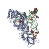
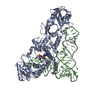
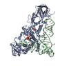
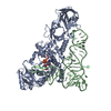
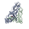
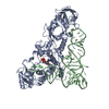
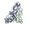
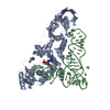
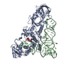
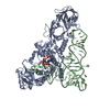
- PDB-4jxx: Crystal structure of E coli E. coli glutaminyl-tRNA synthetase bo... -
+ Open data

- Basic information
| Entry | Database: PDB / ID: 4jxx | ||||||
|---|---|---|---|---|---|---|---|
| Title | Crystal structure of E coli E. coli glutaminyl-tRNA synthetase bound to tRNA(Gln)(CUG) and ATP from novel cryostabilization conditions | ||||||
 Components Components |
| ||||||
 Keywords Keywords | ligase/rna / Rossmann fold / protein-RNA complex / transfer RNA / tRNA aminoacylation / tRNA(Gln) / ligase-rna complex | ||||||
| Function / homology |  Function and homology information Function and homology informationglutamine-tRNA ligase / glutamine-tRNA ligase activity / glutaminyl-tRNA aminoacylation / glutamyl-tRNA aminoacylation / ATP binding / cytosol Similarity search - Function | ||||||
| Biological species |  | ||||||
| Method |  X-RAY DIFFRACTION / SYNCHROTRON / FOURIER SYNTHESIS / Resolution: 2.3 Å X-RAY DIFFRACTION / SYNCHROTRON / FOURIER SYNTHESIS / Resolution: 2.3 Å | ||||||
 Authors Authors | Perona, J.J. / Rodriguez-Hernandez, A. | ||||||
 Citation Citation | Journal: J.Mol.Biol. / Year: 2013 Title: Structural and Mechanistic Basis for Enhanced Translational Efficiency by 2-Thiouridine at the tRNA Anticodon Wobble Position. Authors: Rodriguez-Hernandez, A. / Spears, J.L. / Gaston, K.W. / Limbach, P.A. / Gamper, H. / Hou, Y.M. / Kaiser, R. / Agris, P.F. / Perona, J.J. | ||||||
| History |
|
- Structure visualization
| Structure viewer | Molecule: MolmilJmol/JSmol |
|---|
- Downloads & links
-Download
| PDBx/mmCIF format | 4jxx.cif.gz | 166.4 KB | Display | PDBx/mmCIF format |
|---|---|---|---|---|
| PDB format | pdb4jxx.ent.gz | 125.8 KB | Display | PDB format |
| PDBx/mmJSON format | 4jxx.json.gz | Tree view | PDBx/mmJSON format | |
| Others |  Other downloads Other downloads |
-Validation report
| Arichive directory | https://data.pdbj.org/pub/pdb/validation_reports/jx/4jxxftp://data.pdbj.org/pub/pdb/validation_reports/jx/4jxx | HTTPS FTP |
|---|
-Related structure data
| Related structure data |  4jxzC  4jyzC  1gtrS S: Starting model for refinement C: citing same article ( |
|---|---|
| Similar structure data |
-Links
 PDBj
PDBj


- Assembly
| Deposited unit | 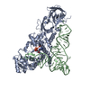
| ||||||||
|---|---|---|---|---|---|---|---|---|---|
| 1 |
| ||||||||
| Unit cell |
| ||||||||
| Components on special symmetry positions |
|
-Components
| #1: Protein | Mass: 63434.641 Da / Num. of mol.: 1 Source method: isolated from a genetically manipulated source Source: (gene. exp.) |
|---|---|
| #2: RNA chain | Mass: 24060.287 Da / Num. of mol.: 1 / Source method: obtained synthetically |
| #3: Chemical | ChemComp-ATP /   Mass: 507.181 Da / Num. of mol.: 1 / Source method: obtained synthetically / Formula: C10H16N5O13P3 / Comment: ATP, energy-carrying molecule*YM Mass: 507.181 Da / Num. of mol.: 1 / Source method: obtained synthetically / Formula: C10H16N5O13P3 / Comment: ATP, energy-carrying molecule*YM |
| #4: Chemical | ChemComp-SO4 /   Mass: 96.063 Da / Num. of mol.: 1 / Source method: obtained synthetically / Formula: SO4 Mass: 96.063 Da / Num. of mol.: 1 / Source method: obtained synthetically / Formula: SO4 |
| #5: Water | ChemComp-HOH /  Mass: 18.015 Da / Num. of mol.: 208 / Source method: isolated from a natural source / Formula: H2O Mass: 18.015 Da / Num. of mol.: 208 / Source method: isolated from a natural source / Formula: H2O |
-Experimental details
-Experiment
| Experiment | Method: X-RAY DIFFRACTION / Number of used crystals: 1 |
|---|
- Sample preparation
| Crystal | Density Matthews: 3.54 Å3/Da / Density % sol: 65.28 % |
|---|---|
| Crystal grow | Temperature: 290 K / Method: vapor diffusion, hanging drop / pH: 7 Details: 0.4 w/v sodium citrate, 40 mM PIPES , 20 mM MgSO4, 20 mM b-mercaptoethanol , pH 7.0, VAPOR DIFFUSION, HANGING DROP, temperature 290.0K PH range: 7.0-7.4 |
-Data collection
| Diffraction | Mean temperature: 100 K |
|---|---|
| Diffraction source | Source: SYNCHROTRON / Site: ALS  / Beamline: 4.2.2 / Wavelength: 1 Å / Beamline: 4.2.2 / Wavelength: 1 Å |
| Detector | Type: NOIR-1 / Detector: CCD / Date: Jan 6, 2012 |
| Radiation | Monochromator: sagitally focused Si(111) / Protocol: SINGLE WAVELENGTH / Monochromatic (M) / Laue (L): M / Scattering type: x-ray |
| Radiation wavelength | Wavelength: 1 Å / Relative weight: 1 |
| Reflection | Resolution: 2.3→41.1 Å / Num. all: 55577 / Num. obs: 55132 / % possible obs: 99.2 % / Observed criterion σ(F): 1 / Observed criterion σ(I): 1 / Redundancy: 7.3 % / Biso Wilson estimate: 50.7 Å2 / Rmerge(I) obs: 0.066 / Net I/σ(I): 12 |
| Reflection shell | Resolution: 2.3→2.38 Å / % possible all: 97.4 |
- Processing
| Software |
| ||||||||||||||||||||
|---|---|---|---|---|---|---|---|---|---|---|---|---|---|---|---|---|---|---|---|---|---|
| Refinement | Method to determine structure: FOURIER SYNTHESIS Starting model: PDB entry 1GTR Resolution: 2.3→41.1 Å / Cross valid method: THROUGHOUT / σ(F): 0 / Stereochemistry target values: Engh & Huber
| ||||||||||||||||||||
| Refine analyze |
| ||||||||||||||||||||
| Refinement step | Cycle: LAST / Resolution: 2.3→41.1 Å
| ||||||||||||||||||||
| Refine LS restraints |
|
