 Movie
Movie Controller

[English] 日本語
 Yorodumi
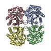
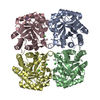
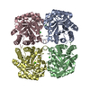
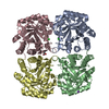
- PDB-4jte: Crystal structure of F114A mutant of 3-deoxy-D-manno-octulosonate... -
+ Open data

- Basic information
| Entry | Database: PDB / ID: 4jte | ||||||
|---|---|---|---|---|---|---|---|
| Title | Crystal structure of F114A mutant of 3-deoxy-D-manno-octulosonate 8-phosphate synthase (KDO8PS) from Neisseria meningitidis | ||||||
 Components Components | 2-dehydro-3-deoxyphosphooctonate aldolase | ||||||
 Keywords Keywords | TRANSFERASE / MANNO-OCTULOSONATE / SYNTHASE / LIPOPOLYSACCHARIDE / KDOP / KDO8 KDOPS / KDO8PS / TIM BARREL / BIOSYNTHESIS / LIPOPOLYSACCHARIDE BIOSYNTHESIS | ||||||
| Function / homology |  Function and homology information Function and homology information3-deoxy-8-phosphooctulonate synthase / 3-deoxy-8-phosphooctulonate synthase activity / keto-3-deoxy-D-manno-octulosonic acid biosynthetic process / cytosol Similarity search - Function | ||||||
| Biological species |  Neisseria meningitidis (bacteria) Neisseria meningitidis (bacteria) | ||||||
| Method |  X-RAY DIFFRACTION / MOLECULAR REPLACEMENT / Resolution: 1.9 Å X-RAY DIFFRACTION / MOLECULAR REPLACEMENT / Resolution: 1.9 Å | ||||||
 Authors Authors | Allison, T.M. / Cochrane, F.C. / Jameson, G.B. / Parker, E.J. | ||||||
 Citation Citation | Journal: Biochemistry / Year: 2013 Title: Examining the Role of Intersubunit Contacts in Catalysis by 3-Deoxy-d-manno-octulosonate 8-Phosphate Synthase. Authors: Allison, T.M. / Cochrane, F.C. / Jameson, G.B. / Parker, E.J. | ||||||
| History |
|
- Structure visualization
| Structure viewer | Molecule: MolmilJmol/JSmol |
|---|
- Downloads & links
-Download
| PDBx/mmCIF format | 4jte.cif.gz | 397.9 KB | Display | PDBx/mmCIF format |
|---|---|---|---|---|
| PDB format | pdb4jte.ent.gz | 328.5 KB | Display | PDB format |
| PDBx/mmJSON format | 4jte.json.gz | Tree view | PDBx/mmJSON format | |
| Others |  Other downloads Other downloads |
-Validation report
| Arichive directory | https://data.pdbj.org/pub/pdb/validation_reports/jt/4jteftp://data.pdbj.org/pub/pdb/validation_reports/jt/4jte | HTTPS FTP |
|---|
-Related structure data
| Related structure data | 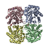 4jtfC 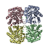 4jtgC  4jthC  4jtiC 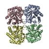 4jtjC  4jtkC  4jtlC 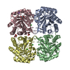 2qkfS C: citing same article ( S: Starting model for refinement |
|---|---|
| Similar structure data |

-Links
 PDBj
PDBj- Assembly
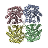
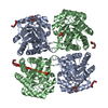
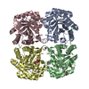
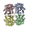
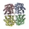
| Deposited unit | 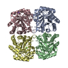
| ||||||||
|---|---|---|---|---|---|---|---|---|---|
| 1 |
| ||||||||
| Unit cell |
|
-Components
| #1: Protein | Mass: 30443.232 Da / Num. of mol.: 4 / Mutation: F114A Source method: isolated from a genetically manipulated source Source: (gene. exp.) Neisseria meningitidis (bacteria) / Strain: MC58 / Gene: kdsA, NMB1283 / Plasmid: pT7-7 / Production host: References: UniProt: Q9JZ55, 3-deoxy-8-phosphooctulonate synthase #2: Chemical | ChemComp-NA / |   Mass: 22.990 Da / Num. of mol.: 1 / Source method: obtained synthetically / Formula: Na Mass: 22.990 Da / Num. of mol.: 1 / Source method: obtained synthetically / Formula: Na#3: Chemical |   Mass: 35.453 Da / Num. of mol.: 2 / Source method: obtained synthetically / Formula: Cl Mass: 35.453 Da / Num. of mol.: 2 / Source method: obtained synthetically / Formula: Cl#4: Water | ChemComp-HOH / |  Mass: 18.015 Da / Num. of mol.: 324 / Source method: isolated from a natural source / Formula: H2O Mass: 18.015 Da / Num. of mol.: 324 / Source method: isolated from a natural source / Formula: H2O |
|---|
-Experimental details
-Experiment
| Experiment | Method: X-RAY DIFFRACTION / Number of used crystals: 1 |
|---|
- Sample preparation
| Crystal | Density Matthews: 2.33 Å3/Da / Density % sol: 47.16 % |
|---|---|
| Crystal grow | Temperature: 297 K / Method: vapor diffusion, hanging drop / pH: 4.6 Details: 20 mg/mL protein (in 10 mM BTP pH 7.5) mixed 1:1 with reservoir liquor containing 100 mM NaOAc (pH 4.6) and 0.6-3.0 M NaCl. Immediately prior to data collection, crystals were harvested and ...Details: 20 mg/mL protein (in 10 mM BTP pH 7.5) mixed 1:1 with reservoir liquor containing 100 mM NaOAc (pH 4.6) and 0.6-3.0 M NaCl. Immediately prior to data collection, crystals were harvested and soaked briefly in cryoprotectant solution, comprising 20% glycerol and the reservoir solution, Vapor diffusion, hanging drop, temperature 297K |
-Data collection
| Diffraction | Mean temperature: 120 K | ||||||||||||||||||||||||||||||||||||||||||||||||||||||||||||||||||||||||||||||||||||||||
|---|---|---|---|---|---|---|---|---|---|---|---|---|---|---|---|---|---|---|---|---|---|---|---|---|---|---|---|---|---|---|---|---|---|---|---|---|---|---|---|---|---|---|---|---|---|---|---|---|---|---|---|---|---|---|---|---|---|---|---|---|---|---|---|---|---|---|---|---|---|---|---|---|---|---|---|---|---|---|---|---|---|---|---|---|---|---|---|---|---|
| Diffraction source | Source: ROTATING ANODE / Type: RIGAKU MICROMAX-007 / Wavelength: 1.5418 Å | ||||||||||||||||||||||||||||||||||||||||||||||||||||||||||||||||||||||||||||||||||||||||
| Detector | Type: RIGAKU RAXIS IV++ / Detector: IMAGE PLATE / Date: Mar 5, 2007 | ||||||||||||||||||||||||||||||||||||||||||||||||||||||||||||||||||||||||||||||||||||||||
| Radiation | Monochromator: AXCO PX70 CAPILLARY OPTIC / Protocol: SINGLE WAVELENGTH / Monochromatic (M) / Laue (L): M / Scattering type: x-ray | ||||||||||||||||||||||||||||||||||||||||||||||||||||||||||||||||||||||||||||||||||||||||
| Radiation wavelength | Wavelength: 1.5418 Å / Relative weight: 1 | ||||||||||||||||||||||||||||||||||||||||||||||||||||||||||||||||||||||||||||||||||||||||
| Reflection | Resolution: 1.9→39.95 Å / Num. obs: 88194 / % possible obs: 97.8 % / Redundancy: 4.7 % / Rmerge(I) obs: 0.065 / Χ2: 0.97 / Net I/σ(I): 8.8 / Scaling rejects: 3133 | ||||||||||||||||||||||||||||||||||||||||||||||||||||||||||||||||||||||||||||||||||||||||
| Reflection shell | Diffraction-ID: 1
|
- Processing
| Software |
| |||||||||||||||||||||||||||||||||||||||||||||||||||||||||||||||||||||||||||||||||||||||||||||||||||||||||||||||||||||||||||||
|---|---|---|---|---|---|---|---|---|---|---|---|---|---|---|---|---|---|---|---|---|---|---|---|---|---|---|---|---|---|---|---|---|---|---|---|---|---|---|---|---|---|---|---|---|---|---|---|---|---|---|---|---|---|---|---|---|---|---|---|---|---|---|---|---|---|---|---|---|---|---|---|---|---|---|---|---|---|---|---|---|---|---|---|---|---|---|---|---|---|---|---|---|---|---|---|---|---|---|---|---|---|---|---|---|---|---|---|---|---|---|---|---|---|---|---|---|---|---|---|---|---|---|---|---|---|---|
| Refinement | Method to determine structure: MOLECULAR REPLACEMENT Starting model: PDB entry 2QKF Resolution: 1.9→39.95 Å / Cor.coef. Fo:Fc: 0.958 / Cor.coef. Fo:Fc free: 0.945 / WRfactor Rfree: 0.2555 / WRfactor Rwork: 0.2175 / Occupancy max: 1 / Occupancy min: 0.5 / FOM work R set: 0.7627 / SU B: 9.734 / SU ML: 0.137 / SU R Cruickshank DPI: 0.1791 / SU Rfree: 0.1632 / Cross valid method: THROUGHOUT / σ(F): 0 / ESU R: 0.179 / ESU R Free: 0.163 / Stereochemistry target values: MAXIMUM LIKELIHOOD Details: U VALUES WITH TLS ADDED HYDROGENS HAVE BEEN ADDED IN THE RIDING POSITIONS
| |||||||||||||||||||||||||||||||||||||||||||||||||||||||||||||||||||||||||||||||||||||||||||||||||||||||||||||||||||||||||||||
| Solvent computation | Ion probe radii: 0.8 Å / Shrinkage radii: 0.8 Å / VDW probe radii: 1.2 Å / Solvent model: MASK | |||||||||||||||||||||||||||||||||||||||||||||||||||||||||||||||||||||||||||||||||||||||||||||||||||||||||||||||||||||||||||||
| Displacement parameters | Biso max: 125.96 Å2 / Biso mean: 46.8224 Å2 / Biso min: 24.39 Å2
| |||||||||||||||||||||||||||||||||||||||||||||||||||||||||||||||||||||||||||||||||||||||||||||||||||||||||||||||||||||||||||||
| Refinement step | Cycle: LAST / Resolution: 1.9→39.95 Å
| |||||||||||||||||||||||||||||||||||||||||||||||||||||||||||||||||||||||||||||||||||||||||||||||||||||||||||||||||||||||||||||
| Refine LS restraints |
| |||||||||||||||||||||||||||||||||||||||||||||||||||||||||||||||||||||||||||||||||||||||||||||||||||||||||||||||||||||||||||||
| LS refinement shell | Resolution: 1.9→1.954 Å / Total num. of bins used: 20
| |||||||||||||||||||||||||||||||||||||||||||||||||||||||||||||||||||||||||||||||||||||||||||||||||||||||||||||||||||||||||||||
| Refinement TLS params. | Method: refined / Refine-ID: X-RAY DIFFRACTION
| |||||||||||||||||||||||||||||||||||||||||||||||||||||||||||||||||||||||||||||||||||||||||||||||||||||||||||||||||||||||||||||
| Refinement TLS group |
|
