 Movie
Movie Controller

[English] 日本語
 Yorodumi
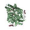
- PDB-4jsd: The X-ray crystal structure of a thermophilic cellobiose binding ... -
+ Open data

- Basic information
| Entry | Database: PDB / ID: 4jsd | |||||||||
|---|---|---|---|---|---|---|---|---|---|---|
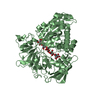
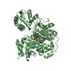
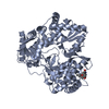
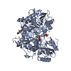
| Title | The X-ray crystal structure of a thermophilic cellobiose binding protein bound with laminaribiose | |||||||||
 Components Components | Oligopeptide ABC transporter, periplasmic oligopeptide-binding protein | |||||||||
 Keywords Keywords | SUGAR BINDING PROTEIN / PERIPLASMIC BINDING PROTEIN / THERMOPHILIC PROTEIN / CELLULOSE / CELLOBIOSE BINDING PROTEIN | |||||||||
| Function / homology |  Function and homology information Function and homology informationpeptide transport / peptide transmembrane transporter activity / metal ion binding Similarity search - Function | |||||||||
| Biological species |   Thermotoga maritima (bacteria) Thermotoga maritima (bacteria) | |||||||||
| Method |  X-RAY DIFFRACTION / MOLECULAR REPLACEMENT / Resolution: 2.05 Å X-RAY DIFFRACTION / MOLECULAR REPLACEMENT / Resolution: 2.05 Å | |||||||||
 Authors Authors | Munshi, P. / Cuneo, M.J. | |||||||||
 Citation Citation | Journal: Bmc Struct.Biol. / Year: 2013 Title: Molecular details of ligand selectivity determinants in a promiscuous beta-glucan periplasmic binding protein. Authors: Munshi, P. / Stanley, C.B. / Ghimire-Rijal, S. / Lu, X. / Myles, D.A. / Cuneo, M.J. | |||||||||
| History |
|
- Structure visualization
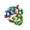
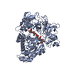
| Structure viewer | Molecule: MolmilJmol/JSmol |
|---|
- Downloads & links
-Download
| PDBx/mmCIF format | 4jsd.cif.gz | 140.6 KB | Display | PDBx/mmCIF format |
|---|---|---|---|---|
| PDB format | pdb4jsd.ent.gz | 107.6 KB | Display | PDB format |
| PDBx/mmJSON format | 4jsd.json.gz | Tree view | PDBx/mmJSON format | |
| Others |  Other downloads Other downloads |
-Validation report
| Arichive directory | https://data.pdbj.org/pub/pdb/validation_reports/js/4jsdftp://data.pdbj.org/pub/pdb/validation_reports/js/4jsd | HTTPS FTP |
|---|
-Related structure data
| Related structure data |  4jsoC  2o7iS C: citing same article ( S: Starting model for refinement |
|---|---|
| Similar structure data |


-Links
 PDBj
PDBj- Assembly
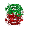
| Deposited unit | 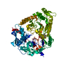
| ||||||||
|---|---|---|---|---|---|---|---|---|---|
| 1 |
| ||||||||
| Unit cell |
|
-Components
| #1: Protein | Mass: 67410.086 Da / Num. of mol.: 1 Source method: isolated from a genetically manipulated source Source: (gene. exp.) Thermotoga maritima (bacteria) / Strain: ATCC 43589 / MSB8 / DSM 3109 / JCM 10099 / Gene: TM0031, TM_0031 / Plasmid: pET21a / Production host: |
|---|---|
| #2: Polysaccharide | beta-D-glucopyranose-(1-3)-beta-D-glucopyranose / beta-laminaribiose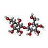  Source method: isolated from a genetically manipulated source Details: oligosaccharide / References: beta-laminaribiose |
| #3: Chemical | ChemComp-CA /   Mass: 40.078 Da / Num. of mol.: 1 / Source method: obtained synthetically / Formula: Ca Mass: 40.078 Da / Num. of mol.: 1 / Source method: obtained synthetically / Formula: Ca |
| #4: Water | ChemComp-HOH /  Mass: 18.015 Da / Num. of mol.: 293 / Source method: isolated from a natural source / Formula: H2O Mass: 18.015 Da / Num. of mol.: 293 / Source method: isolated from a natural source / Formula: H2O |
-Experimental details
-Experiment
| Experiment | Method: X-RAY DIFFRACTION / Number of used crystals: 1 |
|---|
- Sample preparation
| Crystal | Density Matthews: 2.04 Å3/Da / Density % sol: 39.62 % |
|---|---|
| Crystal grow | Temperature: 293 K / Method: vapor diffusion, hanging drop / pH: 8 Details: tmCBP was concentrated to 20 mg/mL and dialyzed into 10 mM Tris, 40 mM NaCl, 0.5 mM TCEP for crystallization. Laminaribiose or laminaripentaose was added to a final concentration of 1 mM ...Details: tmCBP was concentrated to 20 mg/mL and dialyzed into 10 mM Tris, 40 mM NaCl, 0.5 mM TCEP for crystallization. Laminaribiose or laminaripentaose was added to a final concentration of 1 mM prior to crystallization trials. Crystals were grown in drops containing 2 uL of the protein solution mixed with 2 uL of 0.2-0.3 M magnesium acetate or calcium acetate, 20-30% (wt/vol) PEG 3350 equilibrated against 900 uL of the same solution, pH 8.0, VAPOR DIFFUSION, HANGING DROP, temperature 293K |
-Data collection
| Diffraction | Mean temperature: 293 K |
|---|---|
| Diffraction source | Source: ROTATING ANODE / Type: RIGAKU MICROMAX-007 HF / Wavelength: 1.5418 Å |
| Detector | Type: RIGAKU RAXIS IV++ / Detector: IMAGE PLATE / Date: Sep 30, 2012 |
| Radiation | Monochromator: GRAPHITE / Protocol: SINGLE WAVELENGTH / Monochromatic (M) / Laue (L): M / Scattering type: x-ray |
| Radiation wavelength | Wavelength: 1.5418 Å / Relative weight: 1 |
| Reflection | Resolution: 2.05→50 Å / Num. all: 34613 / Num. obs: 34549 / % possible obs: 98.1 % / Observed criterion σ(F): 2 / Observed criterion σ(I): 2 / Redundancy: 3.4 % / Biso Wilson estimate: 30.2 Å2 / Rmerge(I) obs: 0.062 / Rsym value: 0.037 / Net I/σ(I): 15.4 |
| Reflection shell | Resolution: 2.05→2.12 Å / Redundancy: 3.2 % / Rmerge(I) obs: 0.488 / Mean I/σ(I) obs: 2.5 / Num. unique all: 3399 / Rsym value: 0.377 / % possible all: 98.3 |
- Processing
| Software |
| |||||||||||||||||||||||||||||||||||||||||||||||||||||||||||||||||||||||||||||||||||||||||||||||||||||||||
|---|---|---|---|---|---|---|---|---|---|---|---|---|---|---|---|---|---|---|---|---|---|---|---|---|---|---|---|---|---|---|---|---|---|---|---|---|---|---|---|---|---|---|---|---|---|---|---|---|---|---|---|---|---|---|---|---|---|---|---|---|---|---|---|---|---|---|---|---|---|---|---|---|---|---|---|---|---|---|---|---|---|---|---|---|---|---|---|---|---|---|---|---|---|---|---|---|---|---|---|---|---|---|---|---|---|---|
| Refinement | Method to determine structure: MOLECULAR REPLACEMENT Starting model: PDB ENTRY 2O7I Resolution: 2.05→47.876 Å / SU ML: 0.2 / Isotropic thermal model: Isotropic / σ(F): 1.34 / Phase error: 21.74 / Stereochemistry target values: ML
| |||||||||||||||||||||||||||||||||||||||||||||||||||||||||||||||||||||||||||||||||||||||||||||||||||||||||
| Solvent computation | Shrinkage radii: 0.9 Å / VDW probe radii: 1.11 Å / Solvent model: FLAT BULK SOLVENT MODEL | |||||||||||||||||||||||||||||||||||||||||||||||||||||||||||||||||||||||||||||||||||||||||||||||||||||||||
| Refinement step | Cycle: LAST / Resolution: 2.05→47.876 Å
| |||||||||||||||||||||||||||||||||||||||||||||||||||||||||||||||||||||||||||||||||||||||||||||||||||||||||
| Refine LS restraints |
| |||||||||||||||||||||||||||||||||||||||||||||||||||||||||||||||||||||||||||||||||||||||||||||||||||||||||
| LS refinement shell |
|
