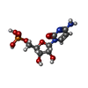
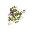
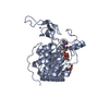
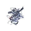
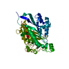
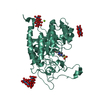
Entry Database : PDB / ID : 4js2Title Crystal structure of human Beta-galactoside alpha-2,6-sialyltransferase 1 in complex with CMP Beta-galactoside alpha-2,6-sialyltransferase 1 Keywords / / / / / / / Function / homology Function Domain/homology Component
/ / / / / / / / / / / / / / / / / / / / / / / / / / / / / / / / / / Biological species Homo sapiens (human)Method / / / Resolution : 2.3 Å Authors Kuhn, B. / Benz, J. / Greif, M. / Engel, A.M. / Sobek, H. / Rudolph, M.G. Journal : Acta Crystallogr.,Sect.D / Year : 2013Title : The structure of human alpha-2,6-sialyltransferase reveals the binding mode of complex glycans.Authors : Kuhn, B. / Benz, J. / Greif, M. / Engel, A.M. / Sobek, H. / Rudolph, M.G. History Deposition Mar 22, 2013 Deposition site / Processing site Revision 1.0 Jul 31, 2013 Provider / Type Revision 1.1 Aug 28, 2013 Group Revision 1.2 Nov 15, 2017 Group / Category Revision 1.3 Dec 18, 2019 Group / Derived calculations / Category / struct_connItem _citation.country / _citation.journal_id_CSD ... _citation.country / _citation.journal_id_CSD / _citation.journal_id_ISSN / _citation.pdbx_database_id_PubMed / _citation.title / _struct_conn.pdbx_leaving_atom_flag Revision 2.0 Jul 29, 2020 Group Advisory / Atomic model ... Advisory / Atomic model / Data collection / Derived calculations / Structure summary Category atom_site / atom_site_anisotrop ... atom_site / atom_site_anisotrop / chem_comp / entity / pdbx_branch_scheme / pdbx_chem_comp_identifier / pdbx_entity_branch / pdbx_entity_branch_descriptor / pdbx_entity_branch_link / pdbx_entity_branch_list / pdbx_entity_nonpoly / pdbx_nonpoly_scheme / pdbx_struct_assembly_gen / pdbx_validate_close_contact / struct_asym / struct_conn / struct_site / struct_site_gen Item _atom_site.auth_asym_id / _atom_site.auth_seq_id ... _atom_site.auth_asym_id / _atom_site.auth_seq_id / _atom_site.label_asym_id / _atom_site.label_entity_id / _atom_site_anisotrop.pdbx_auth_asym_id / _atom_site_anisotrop.pdbx_auth_seq_id / _atom_site_anisotrop.pdbx_label_asym_id / _chem_comp.name / _chem_comp.type / _pdbx_struct_assembly_gen.asym_id_list / _pdbx_validate_close_contact.auth_asym_id_1 / _pdbx_validate_close_contact.auth_asym_id_2 / _pdbx_validate_close_contact.auth_seq_id_1 / _pdbx_validate_close_contact.auth_seq_id_2 / _struct_conn.pdbx_dist_value / _struct_conn.pdbx_leaving_atom_flag / _struct_conn.pdbx_role / _struct_conn.ptnr1_auth_asym_id / _struct_conn.ptnr1_auth_comp_id / _struct_conn.ptnr1_auth_seq_id / _struct_conn.ptnr1_label_asym_id / _struct_conn.ptnr1_label_atom_id / _struct_conn.ptnr1_label_comp_id / _struct_conn.ptnr1_label_seq_id / _struct_conn.ptnr2_auth_asym_id / _struct_conn.ptnr2_auth_comp_id / _struct_conn.ptnr2_auth_seq_id / _struct_conn.ptnr2_label_asym_id / _struct_conn.ptnr2_label_comp_id Description / Provider / Type Revision 2.1 Nov 8, 2023 Group Data collection / Database references ... Data collection / Database references / Derived calculations / Refinement description / Structure summary Category chem_comp / chem_comp_atom ... chem_comp / chem_comp_atom / chem_comp_bond / database_2 / pdbx_initial_refinement_model / struct_conn Item _chem_comp.pdbx_synonyms / _database_2.pdbx_DOI ... _chem_comp.pdbx_synonyms / _database_2.pdbx_DOI / _database_2.pdbx_database_accession / _struct_conn.pdbx_leaving_atom_flag Revision 3.0 May 29, 2024 Group Atomic model / Data collection ... Atomic model / Data collection / Derived calculations / Non-polymer description / Refinement description Category atom_site / atom_site_anisotrop ... atom_site / atom_site_anisotrop / chem_comp / chem_comp_atom / chem_comp_bond / pdbx_entity_nonpoly / pdbx_nonpoly_scheme / pdbx_refine_tls_group Item _atom_site.B_iso_or_equiv / _atom_site.Cartn_x ... _atom_site.B_iso_or_equiv / _atom_site.Cartn_x / _atom_site.Cartn_y / _atom_site.Cartn_z / _atom_site.auth_atom_id / _atom_site.auth_comp_id / _atom_site.label_atom_id / _atom_site.label_comp_id / _atom_site.type_symbol / _atom_site_anisotrop.U[1][1] / _atom_site_anisotrop.U[1][2] / _atom_site_anisotrop.U[1][3] / _atom_site_anisotrop.U[2][2] / _atom_site_anisotrop.U[2][3] / _atom_site_anisotrop.U[3][3] / _atom_site_anisotrop.pdbx_auth_atom_id / _atom_site_anisotrop.pdbx_auth_comp_id / _atom_site_anisotrop.pdbx_label_atom_id / _atom_site_anisotrop.pdbx_label_comp_id / _atom_site_anisotrop.type_symbol / _chem_comp.id / _chem_comp.mon_nstd_flag / _chem_comp.type / _chem_comp_atom.atom_id / _chem_comp_atom.comp_id / _chem_comp_atom.type_symbol / _chem_comp_bond.atom_id_1 / _chem_comp_bond.atom_id_2 / _chem_comp_bond.comp_id / _chem_comp_bond.value_order / _pdbx_entity_nonpoly.comp_id / _pdbx_nonpoly_scheme.mon_id / _pdbx_nonpoly_scheme.pdb_mon_id / _pdbx_refine_tls_group.selection_details Revision 3.1 Nov 20, 2024 Group / Category / pdbx_modification_feature
Show all Show less
 Yorodumi
 Open data
 Basic information
 Components
 Keywords
 Function and homology information
 Homo sapiens (human)
 X-RAY DIFFRACTION /
  SYNCHROTRON /
  MOLECULAR REPLACEMENT / Resolution: 2.3 Å
  Authors
 Citation
 Journal: Acta Crystallogr.,Sect.D / Year: 2013
 Structure visualization
 Molmil
 Jmol/JSmol
 Downloads & links
 Download
 4js2.cif.gz
 PDBx/mmCIF format
 pdb4js2.ent.gz
 PDB format
 4js2.json.gz
 PDBx/mmJSON format
 Other downloads
 https://data.pdbj.org/pub/pdb/validation_reports/js/4js2
 ftp://data.pdbj.org/pub/pdb/validation_reports/js/4js2
 Links
Links Assembly
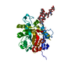
 Components
 Homo sapiens (human) / Gene: ST6GAL1, SIAT1 / Cell line (production host): HEK 293 / Production host:
  Homo sapiens (human) / References: UniProt: P15907, EC: 2.4.99.1
 X-RAY DIFFRACTION / Number of used crystals: 1
  Sample preparation
 SYNCHROTRON / Site:
  SLS
SLS  / Beamline: X10SA / Wavelength: 1 Å
 Processing
 MOLECULAR REPLACEMENT
 Movie
Movie Controller




 PDBj
PDBj