 Movie
Movie Controller

[English] 日本語
 Yorodumi
- PDB-4jr3: Crystal structure of EGFR kinase domain in complex with compound 3g -
+ Open data

- Basic information
| Entry | Database: PDB / ID: 4jr3 | ||||||
|---|---|---|---|---|---|---|---|
| Title | Crystal structure of EGFR kinase domain in complex with compound 3g | ||||||
 Components Components | Epidermal growth factor receptor | ||||||
 Keywords Keywords | TRANSFERASE/TRANSFERASE INHIBITOR / Transferase / tyrosine kinase domain / ATP-binding domain / autophosphorylation / TRANSFERASE-TRANSFERASE INHIBITOR complex | ||||||
| Function / homology |  Function and homology information Function and homology informationmultivesicular body, internal vesicle lumen / negative regulation of cardiocyte differentiation / Shc-EGFR complex / positive regulation of protein kinase C signaling / Inhibition of Signaling by Overexpressed EGFR / epidermal growth factor receptor activity / EGFR interacts with phospholipase C-gamma / regulation of peptidyl-tyrosine phosphorylation / epidermal growth factor binding / response to UV-A ...multivesicular body, internal vesicle lumen / negative regulation of cardiocyte differentiation / Shc-EGFR complex / positive regulation of protein kinase C signaling / Inhibition of Signaling by Overexpressed EGFR / epidermal growth factor receptor activity / EGFR interacts with phospholipase C-gamma / regulation of peptidyl-tyrosine phosphorylation / epidermal growth factor binding / response to UV-A / PLCG1 events in ERBB2 signaling / ERBB2-EGFR signaling pathway / morphogenesis of an epithelial fold / PTK6 promotes HIF1A stabilization / ERBB2 Activates PTK6 Signaling / digestive tract morphogenesis / Signaling by EGFR / intracellular vesicle / negative regulation of epidermal growth factor receptor signaling pathway / eyelid development in camera-type eye / cerebral cortex cell migration / protein insertion into membrane / ERBB2 Regulates Cell Motility / protein tyrosine kinase activator activity / Respiratory syncytial virus (RSV) attachment and entry / Signaling by ERBB4 / PI3K events in ERBB2 signaling / positive regulation of phosphorylation / positive regulation of peptidyl-serine phosphorylation / Estrogen-dependent nuclear events downstream of ESR-membrane signaling / hair follicle development / MAP kinase kinase kinase activity / GAB1 signalosome / positive regulation of G1/S transition of mitotic cell cycle / embryonic placenta development / salivary gland morphogenesis / Signaling by ERBB2 / TFAP2 (AP-2) family regulates transcription of growth factors and their receptors / GRB2 events in EGFR signaling / SHC1 events in EGFR signaling / transmembrane receptor protein tyrosine kinase activity / EGFR Transactivation by Gastrin / GRB2 events in ERBB2 signaling / ossification / SHC1 events in ERBB2 signaling / basal plasma membrane / positive regulation of DNA repair / cellular response to epidermal growth factor stimulus / positive regulation of DNA replication / epithelial cell proliferation / positive regulation of epithelial cell proliferation / Signal transduction by L1 / positive regulation of protein localization to plasma membrane / NOTCH3 Activation and Transmission of Signal to the Nucleus / cellular response to amino acid stimulus / phosphatidylinositol 3-kinase/protein kinase B signal transduction / cellular response to estradiol stimulus / EGFR downregulation / clathrin-coated endocytic vesicle membrane / Signaling by ERBB2 TMD/JMD mutants / Constitutive Signaling by EGFRvIII / cell-cell adhesion / receptor protein-tyrosine kinase / Signaling by ERBB2 ECD mutants / negative regulation of protein catabolic process / Signaling by ERBB2 KD Mutants / positive regulation of miRNA transcription / kinase binding / ruffle membrane / Downregulation of ERBB2 signaling / epidermal growth factor receptor signaling pathway / positive regulation of protein phosphorylation / cell morphogenesis / positive regulation of fibroblast proliferation / neuron differentiation / HCMV Early Events / Constitutive Signaling by Aberrant PI3K in Cancer / actin filament binding / cell junction / transmembrane signaling receptor activity / positive regulation of canonical Wnt signaling pathway / Cargo recognition for clathrin-mediated endocytosis / PIP3 activates AKT signaling / Constitutive Signaling by Ligand-Responsive EGFR Cancer Variants / Clathrin-mediated endocytosis / virus receptor activity / ATPase binding / PI5P, PP2A and IER3 Regulate PI3K/AKT Signaling / RAF/MAP kinase cascade / positive regulation of cell growth / double-stranded DNA binding / protein tyrosine kinase activity / early endosome membrane / protein phosphatase binding / nuclear membrane / basolateral plasma membrane / learning or memory / Extra-nuclear estrogen signaling / cell surface receptor signaling pathway / positive regulation of ERK1 and ERK2 cascade Similarity search - Function | ||||||
| Biological species |  Homo sapiens (human) Homo sapiens (human) | ||||||
| Method |  X-RAY DIFFRACTION / SYNCHROTRON / MOLECULAR REPLACEMENT / Resolution: 2.7 Å X-RAY DIFFRACTION / SYNCHROTRON / MOLECULAR REPLACEMENT / Resolution: 2.7 Å | ||||||
 Authors Authors | Peng, Y.H. / Wu, J.S. | ||||||
 Citation Citation | Journal: J.Med.Chem. / Year: 2013 Title: Protein Kinase Inhibitor Design by Targeting the Asp-Phe-Gly (DFG) Motif: The Role of the DFG Motif in the Design of Epidermal Growth Factor Receptor Inhibitors Authors: Peng, Y.H. / Shiao, H.Y. / Tu, C.H. / Liu, P.M. / Hsu, J.T. / Amancha, P.K. / Wu, J.S. / Coumar, M.S. / Chen, C.H. / Wang, S.Y. / Lin, W.H. / Sun, H.Y. / Chao, Y.S. / Lyu, P.C. / Hsieh, H.P. / Wu, S.Y. | ||||||
| History |
|
- Structure visualization
| Structure viewer | Molecule: MolmilJmol/JSmol |
|---|
- Downloads & links
-Download
| PDBx/mmCIF format | 4jr3.cif.gz | 137.3 KB | Display | PDBx/mmCIF format |
|---|---|---|---|---|
| PDB format | pdb4jr3.ent.gz | 107.6 KB | Display | PDB format |
| PDBx/mmJSON format | 4jr3.json.gz | Tree view | PDBx/mmJSON format | |
| Others |  Other downloads Other downloads |
-Validation report
| Summary document | 4jr3_validation.pdf.gz | 811 KB | Display | wwPDB validaton report |
|---|---|---|---|---|
| Full document | 4jr3_full_validation.pdf.gz | 814.1 KB | Display | |
| Data in XML | 4jr3_validation.xml.gz | 13.2 KB | Display | |
| Data in CIF | 4jr3_validation.cif.gz | 17.2 KB | Display | |
| Arichive directory | https://data.pdbj.org/pub/pdb/validation_reports/jr/4jr3ftp://data.pdbj.org/pub/pdb/validation_reports/jr/4jr3 | HTTPS FTP |
-Related structure data
| Related structure data |  4jq7C  4jq8C  4jrvC  1m17S C: citing same article ( S: Starting model for refinement |
|---|---|
| Similar structure data |


-Links
 PDBj
PDBj


- Assembly
| Deposited unit | 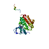
| ||||||||
|---|---|---|---|---|---|---|---|---|---|
| 1 | 
| ||||||||
| Unit cell |
|
-Components
| #1: Protein | Mass: 37401.250 Da / Num. of mol.: 1 / Fragment: EGFR kinase domain, UNP residues 696-1021 Source method: isolated from a genetically manipulated source Source: (gene. exp.) Homo sapiens (human) / Gene: EGFR / Plasmid: pBacPAK-MT-EGFP / Cell line (production host): Hi5 / Production host:   Spodoptera frugiperda (fall armyworm) Spodoptera frugiperda (fall armyworm)References: UniProt: P00533, receptor protein-tyrosine kinase |
|---|---|
| #2: Chemical | ChemComp-KJR /   Mass: 464.515 Da / Num. of mol.: 1 / Source method: obtained synthetically / Formula: C28H24N4O3 Mass: 464.515 Da / Num. of mol.: 1 / Source method: obtained synthetically / Formula: C28H24N4O3 |
| #3: Water | ChemComp-HOH /  Mass: 18.015 Da / Num. of mol.: 20 / Source method: isolated from a natural source / Formula: H2O Mass: 18.015 Da / Num. of mol.: 20 / Source method: isolated from a natural source / Formula: H2O |
-Experimental details
-Experiment
| Experiment | Method: X-RAY DIFFRACTION / Number of used crystals: 1 |
|---|
- Sample preparation
| Crystal | Density Matthews: 3.44 Å3/Da / Density % sol: 64.3 % |
|---|---|
| Crystal grow | Temperature: 291 K / Method: vapor diffusion, hanging drop / pH: 7 Details: 1.0M Ammonium citrate tribase, 0.1M Bis-Tris propane, pH 7.0, VAPOR DIFFUSION, HANGING DROP, temperature 291K |
-Data collection
| Diffraction | Mean temperature: 100 K |
|---|---|
| Diffraction source | Source: SYNCHROTRON / Site: SPring-8  / Beamline: BL12B2 / Wavelength: 1 Å / Beamline: BL12B2 / Wavelength: 1 Å |
| Detector | Type: ADSC QUANTUM 210 / Detector: CCD / Date: May 25, 2012 |
| Radiation | Protocol: SINGLE WAVELENGTH / Monochromatic (M) / Laue (L): M / Scattering type: x-ray |
| Radiation wavelength | Wavelength: 1 Å / Relative weight: 1 |
| Reflection | Resolution: 2.7→30 Å / Num. all: 14311 / Num. obs: 14311 / % possible obs: 99.4 % / Observed criterion σ(F): 0 / Observed criterion σ(I): 0 / Redundancy: 10.5 % / Rmerge(I) obs: 0.071 / Net I/σ(I): 19.92 |
| Reflection shell | Resolution: 2.7→2.8 Å / Redundancy: 4.5 % / Rmerge(I) obs: 0.468 / Mean I/σ(I) obs: 3.2 / Num. unique all: 1399 / % possible all: 100 |
- Processing
| Software |
| ||||||||||||||||||||||||||||||||||||||||||||||||||||||||||||
|---|---|---|---|---|---|---|---|---|---|---|---|---|---|---|---|---|---|---|---|---|---|---|---|---|---|---|---|---|---|---|---|---|---|---|---|---|---|---|---|---|---|---|---|---|---|---|---|---|---|---|---|---|---|---|---|---|---|---|---|---|---|
| Refinement | Method to determine structure: MOLECULAR REPLACEMENT Starting model: PDB entry 1M17 Resolution: 2.7→30 Å / Cor.coef. Fo:Fc: 0.933 / Cor.coef. Fo:Fc free: 0.917 / SU B: 22.491 / SU ML: 0.214 / Cross valid method: THROUGHOUT / ESU R: 0.484 / ESU R Free: 0.292 / Stereochemistry target values: MAXIMUM LIKELIHOOD / Details: HYDROGENS HAVE BEEN ADDED IN THE RIDING POSITIONS
| ||||||||||||||||||||||||||||||||||||||||||||||||||||||||||||
| Solvent computation | Ion probe radii: 0.8 Å / Shrinkage radii: 0.8 Å / VDW probe radii: 1.1 Å / Solvent model: MASK | ||||||||||||||||||||||||||||||||||||||||||||||||||||||||||||
| Displacement parameters | Biso mean: 61.385 Å2
| ||||||||||||||||||||||||||||||||||||||||||||||||||||||||||||
| Refinement step | Cycle: LAST / Resolution: 2.7→30 Å
| ||||||||||||||||||||||||||||||||||||||||||||||||||||||||||||
| Refine LS restraints |
| ||||||||||||||||||||||||||||||||||||||||||||||||||||||||||||
| LS refinement shell | Resolution: 2.7→2.77 Å / Total num. of bins used: 20
| ||||||||||||||||||||||||||||||||||||||||||||||||||||||||||||
| Refinement TLS params. | Method: refined / Origin x: 58.445 Å / Origin y: 23.961 Å / Origin z: 9.65 Å
|
