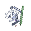
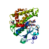
CUE1-UBC7 ubiquitin-conjugating enzyme complex / establishment of protein localization to endoplasmic reticulum membrane / Doa10p ubiquitin ligase complex / Synthesis of active ubiquitin: roles of E1 and E2 enzymes / Hrd1p ubiquitin ligase ERAD-L complex / fungal-type cell wall organization / ubiquitin-protein transferase activator activity / E2 ubiquitin-conjugating enzyme / ubiquitin conjugating enzyme activity / Antigen processing: Ubiquitination & Proteasome degradation ...CUE1-UBC7 ubiquitin-conjugating enzyme complex / establishment of protein localization to endoplasmic reticulum membrane / Doa10p ubiquitin ligase complex / Synthesis of active ubiquitin: roles of E1 and E2 enzymes / Hrd1p ubiquitin ligase ERAD-L complex / fungal-type cell wall organization / ubiquitin-protein transferase activator activity / E2 ubiquitin-conjugating enzyme / ubiquitin conjugating enzyme activity / Antigen processing: Ubiquitination & Proteasome degradation / response to cadmium ion / ERAD pathway / ubiquitin binding / protein polyubiquitination / ubiquitin-protein transferase activity / chromatin organization / ubiquitin-dependent protein catabolic process / endoplasmic reticulum membrane / endoplasmic reticulum / mitochondrion / ATP binding Similarity search - Function
Helix Hairpins - #4310 / Cue1, Ubc7p-binding domain / Ubc7p-binding region of Cue1 / CUE domain / Domain that may be involved in binding ubiquitin-conjugating enzymes (UBCs) / Ubiquitin system component CUE / CUE domain profile. / : / Ubiquitin Conjugating Enzyme / Ubiquitin Conjugating Enzyme ...Helix Hairpins - #4310 / Cue1, Ubc7p-binding domain / Ubc7p-binding region of Cue1 / CUE domain / Domain that may be involved in binding ubiquitin-conjugating enzymes (UBCs) / Ubiquitin system component CUE / CUE domain profile. / : / Ubiquitin Conjugating Enzyme / Ubiquitin Conjugating Enzyme / Ubiquitin-conjugating enzyme, active site / Ubiquitin-conjugating (UBC) active site signature. / Ubiquitin-conjugating enzyme E2 / Ubiquitin-conjugating enzyme / Ubiquitin-conjugating (UBC) core domain profile. / Ubiquitin-conjugating enzyme E2, catalytic domain homologues / Ubiquitin-conjugating enzyme/RWD-like / Helix Hairpins / Roll / Orthogonal Bundle / Mainly Alpha / Alpha Beta Similarity search - Domain/homology
DI(HYDROXYETHYL)ETHER / Coupling of ubiquitin conjugation to ER degradation protein 1 / Ubiquitin-conjugating enzyme E2 7 Similarity search - Component
In the structure databanks used in Yorodumi, some data are registered as the other names, "COVID-19 virus" and "2019-nCoV". Here are the details of the virus and the list of structure data.
Jan 31, 2019. EMDB accession codes are about to change! (news from PDBe EMDB page)
EMDB accession codes are about to change! (news from PDBe EMDB page)
The allocation of 4 digits for EMDB accession codes will soon come to an end. Whilst these codes will remain in use, new EMDB accession codes will include an additional digit and will expand incrementally as the available range of codes is exhausted. The current 4-digit format prefixed with “EMD-” (i.e. EMD-XXXX) will advance to a 5-digit format (i.e. EMD-XXXXX), and so on. It is currently estimated that the 4-digit codes will be depleted around Spring 2019, at which point the 5-digit format will come into force.
The EM Navigator/Yorodumi systems omit the EMD- prefix.
Related info.:Q: What is EMD? / ID/Accession-code notation in Yorodumi/EM Navigator
Yorodumi is a browser for structure data from EMDB, PDB, SASBDB, etc.
This page is also the successor to EM Navigator detail page, and also detail information page/front-end page for Omokage search.
The word "yorodu" (or yorozu) is an old Japanese word meaning "ten thousand". "mi" (miru) is to see.
Related info.:EMDB / PDB / SASBDB / Comparison of 3 databanks / Yorodumi Search / Aug 31, 2016. New EM Navigator & Yorodumi / Yorodumi Papers / Jmol/JSmol / Function and homology information / Changes in new EM Navigator and Yorodumi
 Movie
Movie Controller


 Open data
 Basic information
 Components
 Keywords
 Function and homology information
 X-RAY DIFFRACTION /
  Authors
 Citation
 Structure visualization
 Downloads & links
 Other downloads


 PDBj
PDBj


 Assembly
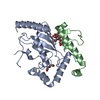


 Mass: 209.240 Da / Num. of mol.: 1 / Source method: obtained synthetically / Formula: C8H19NO5 / Comment: pH buffer*YM
 Mass: 106.120 Da / Num. of mol.: 2 / Source method: obtained synthetically / Formula: C4H10O3
 Mass: 18.015 Da / Num. of mol.: 136 / Source method: isolated from a natural source / Formula: H2O
 Sample preparation
 / Beamline: 22-BM / Wavelength: 1 Å
 Processing