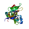
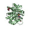
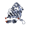
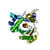
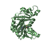
- PDB-4jqr: Crystal structure of a DUF4465 family protein (BACCAC_02373) from... -
+
Open data
ID or keywords:
Loading...
-
Basic information
Entry
Database: PDB / ID: 4jqr
Title
Crystal structure of a DUF4465 family protein (BACCAC_02373) from Bacteroides caccae ATCC 43185 at 2.05 A resolution
Components
hypothetical protein
Keywords
Structural Genomics / Unknown Function / PF14717 family protein / DUF4465 / Joint Center for Structural Genomics / JCSG / Protein Structure Initiative / PSI-BIOLOGY
Function / homology
Protein of unknown function DUF4465 / Protein of unknown function DUF4465 / Domain of unknown function (DUF4465) / Jelly Rolls / Sandwich / Mainly Beta / Uncharacterized protein
Function and homology information
Biological species
Bacteroides caccae (bacteria)
Method
X-RAY DIFFRACTION / SYNCHROTRON / MAD / Resolution: 2.05 Å
Mass: 18.015 Da / Num. of mol.: 258 / Source method: isolated from a natural source / Formula: H2O
-
Details
Has protein modification
Y
Sequence details
THE CONSTRUCT (20-247) WAS EXPRESSED WITH A PURIFICATION TAG MGSDKIHHHHHHENLYFQG. THE TAG WAS ...THE CONSTRUCT (20-247) WAS EXPRESSED WITH A PURIFICATION TAG MGSDKIHHHHHHENLYFQG. THE TAG WAS REMOVED WITH TEV PROTEASE LEAVING ONLY A GLYCINE (0) FOLLOWED BY THE TARGET SEQUENCE.
-
Experimental details
-
Experiment
Experiment
Method: X-RAY DIFFRACTION / Number of used crystals: 1
-
Sample preparation
Crystal
Density Matthews: 3.79 Å3/Da / Density % sol: 67.56 %
Resolution: 2.05→29.249 Å / Num. all: 24729 / Num. obs: 24729 / % possible obs: 100 % / Redundancy: 7.2 % / Rsym value: 0.131 / Net I/σ(I): 10.7
Reflection shell
Rmerge(I) obs: 0.013 / Diffraction-ID: 1
Resolution (Å)
Redundancy (%)
Mean I/σ(I) obs
Num. measured all
Num. unique all
Rsym value
% possible all
2.05-2.1
7.3
1.8
13114
1803
1.328
100
2.1-2.16
7.3
2.4
12773
1746
0.98
100
2.16-2.22
7.3
2.7
12585
1718
0.851
100
2.22-2.29
7.3
3.2
12198
1675
0.728
100
2.29-2.37
7.3
4
11864
1624
0.568
100
2.37-2.45
7.3
4.4
11338
1555
0.501
100
2.45-2.54
7.3
5.2
10864
1495
0.408
100
2.54-2.65
7.2
6
10652
1470
0.343
100
2.65-2.76
7.2
7.4
10054
1396
0.26
100
2.76-2.9
7.2
9.4
9739
1347
0.192
100
2.9-3.06
7.2
11.9
9069
1266
0.146
100
3.06-3.24
7.2
14.9
8677
1209
0.113
100
3.24-3.47
7.1
18.9
8162
1152
0.086
100
3.47-3.74
7
22.4
7461
1059
0.069
100
3.74-4.1
6.9
24.8
6822
993
0.058
100
4.1-4.58
6.7
27.5
5989
891
0.054
100
4.58-5.29
6.4
27.9
5146
803
0.059
100
5.29-6.48
7.2
26.5
4828
671
0.068
100
6.48-9.17
7.1
28.8
3865
546
0.045
100
9.17-29.249
6.6
30.8
2036
310
0.034
97.4
-
Phasing
Phasing
Method: MAD
-
Processing
Software
Name
Version
Classification
NB
MolProbity
3beta29
modelbuilding
PDB_EXTRACT
3.1
dataextraction
SHELX
phasing
SHARP
phasing
SCALA
3.3.20
datascaling
BUSTER-TNT
2.10.0
refinement
MOSFLM
datareduction
SHELXD
phasing
BUSTER
2.10.0
refinement
Refinement
Method to determine structure: MAD / Resolution: 2.05→29.249 Å / Cor.coef. Fo:Fc: 0.9511 / Cor.coef. Fo:Fc free: 0.944 / Occupancy max: 1 / Occupancy min: 0.5 / Cross valid method: THROUGHOUT / σ(F): 0 Details: 1. A MET-INHIBITION PROTOCOL WAS USED FOR SELENOMETHIONINE INCORPORATION DURING PROTEIN EXPRESSION. THE OCCUPANCY OF THE SE ATOMS IN THE MSE RESIDUES WAS REDUCED TO 0.75 FOR THE REDUCED ...Details: 1. A MET-INHIBITION PROTOCOL WAS USED FOR SELENOMETHIONINE INCORPORATION DURING PROTEIN EXPRESSION. THE OCCUPANCY OF THE SE ATOMS IN THE MSE RESIDUES WAS REDUCED TO 0.75 FOR THE REDUCED SCATTERING POWER DUE TO PARTIAL S-MET INCORPORATION. 2. ATOM RECORD CONTAINS SUM OF TLS AND RESIDUAL B FACTORS. ANISOU RECORD CONTAINS SUM OF TLS AND RESIDUAL U FACTORS. 3. THE MAD PHASES WERE USED AS RESTRAINTS DURING REFINEMENT. 4. NA, CL, SO4 AND EDO ARE PRESENT IN CRYSTALLIZATION/CRYO CONDITIONS.
In the structure databanks used in Yorodumi, some data are registered as the other names, "COVID-19 virus" and "2019-nCoV". Here are the details of the virus and the list of structure data.
Jan 31, 2019. EMDB accession codes are about to change! (news from PDBe EMDB page)
EMDB accession codes are about to change! (news from PDBe EMDB page)
The allocation of 4 digits for EMDB accession codes will soon come to an end. Whilst these codes will remain in use, new EMDB accession codes will include an additional digit and will expand incrementally as the available range of codes is exhausted. The current 4-digit format prefixed with “EMD-” (i.e. EMD-XXXX) will advance to a 5-digit format (i.e. EMD-XXXXX), and so on. It is currently estimated that the 4-digit codes will be depleted around Spring 2019, at which point the 5-digit format will come into force.
The EM Navigator/Yorodumi systems omit the EMD- prefix.
Related info.:Q: What is EMD? / ID/Accession-code notation in Yorodumi/EM Navigator
Yorodumi is a browser for structure data from EMDB, PDB, SASBDB, etc.
This page is also the successor to EM Navigator detail page, and also detail information page/front-end page for Omokage search.
The word "yorodu" (or yorozu) is an old Japanese word meaning "ten thousand". "mi" (miru) is to see.
Related info.:EMDB / PDB / SASBDB / Comparison of 3 databanks / Yorodumi Search / Aug 31, 2016. New EM Navigator & Yorodumi / Yorodumi Papers / Jmol/JSmol / Function and homology information / Changes in new EM Navigator and Yorodumi
 Movie
Movie Controller

 Yorodumi
 Open data
 Basic information
 Components
 Keywords
 Function and homology information
 Bacteroides caccae (bacteria)
 X-RAY DIFFRACTION /
  Authors
 Citation
 Structure visualization
 Downloads & links
 Other downloads


 PDBj
PDBj Assembly
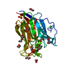


 Mass: 22.990 Da / Num. of mol.: 1 / Source method: obtained synthetically / Formula: Na
 Mass: 35.453 Da / Num. of mol.: 1 / Source method: obtained synthetically / Formula: Cl
 Mass: 96.063 Da / Num. of mol.: 1 / Source method: obtained synthetically / Formula: SO4
 Mass: 62.068 Da / Num. of mol.: 15 / Source method: obtained synthetically / Formula: C2H6O2
 Sample preparation
 / Beamline: 8.2.2 / Wavelength: 0.918374,0.979415,0.979129
 Processing