Protocol: SINGLE WAVELENGTH / Scattering type: x-ray
Radiation wavelength
Wavelength: 0.9791 Å / Relative weight: 1
Reflection
Redundancy: 7 % / Av σ(I) over netI: 16.91 / Number: 166427 / Rmerge(I) obs: 0.095 / Χ2: 1.04 / D res high: 2.2 Å / D res low: 50 Å / Num. obs: 23688 / % possible obs: 99.9
Diffraction reflection shell
Highest resolution (Å)
Lowest resolution (Å)
% possible obs (%)
ID
Rmerge(I) obs
Chi squared
Redundancy
4.74
50
99.2
1
0.066
1.092
6
3.76
4.74
99.8
1
0.06
1.086
6.6
3.29
3.76
99.9
1
0.071
1.063
6.9
2.99
3.29
99.9
1
0.093
1.004
7.1
2.77
2.99
100
1
0.122
1.03
7.2
2.61
2.77
100
1
0.158
1.058
7.3
2.48
2.61
100
1
0.209
1.063
7.3
2.37
2.48
100
1
0.273
1.071
7.3
2.28
2.37
100
1
0.353
0.986
7.3
2.2
2.28
100
1
0.457
0.988
7.3
Reflection
Resolution: 2.2→50 Å / Num. obs: 23688 / % possible obs: 99.9 % / Redundancy: 7 % / Biso Wilson estimate: 28.1 Å2 / Rmerge(I) obs: 0.095 / Rrim(I) all: 0.095 / Χ2: 1.043 / Net I/σ(I): 16.907
Reflection shell
Resolution (Å)
Redundancy (%)
Rmerge(I) obs
Num. unique all
Χ2
Diffraction-ID
% possible all
2.2-2.28
7.3
0.457
2326
0.988
1
100
2.28-2.37
7.3
0.353
2327
0.986
1
100
2.37-2.48
7.3
0.273
2334
1.071
1
100
2.48-2.61
7.3
0.209
2330
1.063
1
100
2.61-2.77
7.3
0.158
2351
1.058
1
100
2.77-2.99
7.2
0.122
2336
1.03
1
100
2.99-3.29
7.1
0.093
2391
1.004
1
99.9
3.29-3.76
6.9
0.071
2369
1.063
1
99.9
3.76-4.74
6.6
0.06
2397
1.086
1
99.8
4.74-50
6
0.066
2527
1.092
1
99.2
-
Processing
Software
Name
Version
Classification
NB
DENZO
datareduction
SCALEPACK
datascaling
PHENIX
1.7_650
refinement
PDB_EXTRACT
3.11
dataextraction
HKL-2000
datacollection
HKL-2000
datareduction
HKL-2000
datascaling
AutoSol
phasing
Refinement
Method to determine structure: SAD / Resolution: 2.21→46.39 Å / Occupancy max: 1 / Occupancy min: 1 / FOM work R set: 0.8817 / SU ML: 0.18 / σ(F): 0 / Phase error: 18.18 / Stereochemistry target values: ML
Rfactor
Num. reflection
% reflection
Selection details
Rfree
0.201
1172
5.11 %
RANDOM
Rwork
0.1688
21746
-
-
obs
0.1704
22918
96.41 %
-
Solvent computation
Shrinkage radii: 0.83 Å / VDW probe radii: 1.1 Å / Solvent model: FLAT BULK SOLVENT MODEL / Bsol: 40.643 Å2 / ksol: 0.351 e/Å3
In the structure databanks used in Yorodumi, some data are registered as the other names, "COVID-19 virus" and "2019-nCoV". Here are the details of the virus and the list of structure data.
Jan 31, 2019. EMDB accession codes are about to change! (news from PDBe EMDB page)
EMDB accession codes are about to change! (news from PDBe EMDB page)
The allocation of 4 digits for EMDB accession codes will soon come to an end. Whilst these codes will remain in use, new EMDB accession codes will include an additional digit and will expand incrementally as the available range of codes is exhausted. The current 4-digit format prefixed with “EMD-” (i.e. EMD-XXXX) will advance to a 5-digit format (i.e. EMD-XXXXX), and so on. It is currently estimated that the 4-digit codes will be depleted around Spring 2019, at which point the 5-digit format will come into force.
The EM Navigator/Yorodumi systems omit the EMD- prefix.
Related info.:Q: What is EMD? / ID/Accession-code notation in Yorodumi/EM Navigator
Yorodumi is a browser for structure data from EMDB, PDB, SASBDB, etc.
This page is also the successor to EM Navigator detail page, and also detail information page/front-end page for Omokage search.
The word "yorodu" (or yorozu) is an old Japanese word meaning "ten thousand". "mi" (miru) is to see.
Related info.:EMDB / PDB / SASBDB / Comparison of 3 databanks / Yorodumi Search / Aug 31, 2016. New EM Navigator & Yorodumi / Yorodumi Papers / Jmol/JSmol / Function and homology information / Changes in new EM Navigator and Yorodumi
 Movie
Movie Controller

 Yorodumi
 Open data
 Basic information
 Components
 Keywords
 Function and homology information
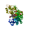
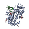
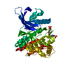
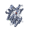
 Homo sapiens (human)
 X-RAY DIFFRACTION /
  Authors
 Citation
 Structure visualization
 Downloads & links
 Other downloads


 PDBj
PDBj


 Assembly
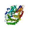


 Mass: 122.143 Da / Num. of mol.: 1 / Source method: obtained synthetically / Formula: C4H12NO3 / Comment: pH buffer*YM
 Mass: 18.015 Da / Num. of mol.: 136 / Source method: isolated from a natural source / Formula: H2O
 Sample preparation
 / Beamline: BL-17A / Wavelength: 0.9791 Å
 Processing