 Movie
Movie Controller

[English] 日本語
 Yorodumi
- PDB-4jo8: Crystal structure of the activating Ly49H receptor in complex wit... -
+ Open data

- Basic information
| Entry | Database: PDB / ID: 4jo8 | ||||||
|---|---|---|---|---|---|---|---|
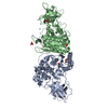
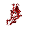
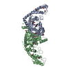
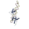
| Title | Crystal structure of the activating Ly49H receptor in complex with m157 (G1F strain) | ||||||
 Components Components |
| ||||||
 Keywords Keywords | IMMUNE SYSTEM/VIRAL PROTEIN / C-type lectin-like domain / natural killer receptor / viral immunoevasin / IMMUNE SYSTEM-VIRAL PROTEIN complex | ||||||
| Function / homology |  Function and homology information Function and homology informationresponse to virus / carbohydrate binding / cell adhesion / cell surface / plasma membrane Similarity search - Function | ||||||
| Biological species |  Murid herpesvirus 1 (Murine cytomegalovirus) Murid herpesvirus 1 (Murine cytomegalovirus) | ||||||
| Method |  X-RAY DIFFRACTION / SYNCHROTRON / MOLECULAR REPLACEMENT / Resolution: 3.2 Å X-RAY DIFFRACTION / SYNCHROTRON / MOLECULAR REPLACEMENT / Resolution: 3.2 Å | ||||||
 Authors Authors | Berry, R. / Ng, N. / Saunders, P.M. / Vivian, J.P. / Lin, J. / Deuss, F.A. / Corbett, A.J. / Forbes, C.A. / Widjaja, J.M. / Sullivan, L.C. ...Berry, R. / Ng, N. / Saunders, P.M. / Vivian, J.P. / Lin, J. / Deuss, F.A. / Corbett, A.J. / Forbes, C.A. / Widjaja, J.M. / Sullivan, L.C. / McAlister, A.D. / Perugini, M.A. / Call, M.J. / Scalzo, A.A. / Degli-Esposti, M.A. / Coudert, J.D. / Beddoe, T. / Brooks, A.G. / Rossjohn, J. | ||||||
 Citation Citation | Journal: Nat.Immunol. / Year: 2013 Title: Targeting of a natural killer cell receptor family by a viral immunoevasin Authors: Berry, R. / Ng, N. / Saunders, P.M. / Vivian, J.P. / Lin, J. / Deuss, F.A. / Corbett, A.J. / Forbes, C.A. / Widjaja, J.M. / Sullivan, L.C. / McAlister, A.D. / Perugini, M.A. / Call, M.J. / ...Authors: Berry, R. / Ng, N. / Saunders, P.M. / Vivian, J.P. / Lin, J. / Deuss, F.A. / Corbett, A.J. / Forbes, C.A. / Widjaja, J.M. / Sullivan, L.C. / McAlister, A.D. / Perugini, M.A. / Call, M.J. / Scalzo, A.A. / Degli-Esposti, M.A. / Coudert, J.D. / Beddoe, T. / Brooks, A.G. / Rossjohn, J. | ||||||
| History |
|
- Structure visualization
| Structure viewer | Molecule: MolmilJmol/JSmol |
|---|
- Downloads & links
-Download
| PDBx/mmCIF format | 4jo8.cif.gz | 127.2 KB | Display | PDBx/mmCIF format |
|---|---|---|---|---|
| PDB format | pdb4jo8.ent.gz | 97.3 KB | Display | PDB format |
| PDBx/mmJSON format | 4jo8.json.gz | Tree view | PDBx/mmJSON format | |
| Others |  Other downloads Other downloads |
-Validation report
| Arichive directory | https://data.pdbj.org/pub/pdb/validation_reports/jo/4jo8ftp://data.pdbj.org/pub/pdb/validation_reports/jo/4jo8 | HTTPS FTP |
|---|
-Related structure data
| Related structure data |  2nykS S: Starting model for refinement |
|---|---|
| Similar structure data |


-Links
 PDBj
PDBj


- Assembly
| Deposited unit | 
| ||||||||
|---|---|---|---|---|---|---|---|---|---|
| 1 | 
| ||||||||
| Unit cell |
|
-Components
| #1: Protein | Mass: 30375.441 Da / Num. of mol.: 1 / Fragment: UNP residues 29-291 / Mutation: P29A, D30G Source method: isolated from a genetically manipulated source Source: (gene. exp.) Murid herpesvirus 1 (Murine cytomegalovirus)Gene: m157 / Plasmid: PHLSEC / Cell line (production host): HEK293S / Production host:  HOMO SAPIENS (human) / References: UniProt: Q6XK91 HOMO SAPIENS (human) / References: UniProt: Q6XK91 |
|---|---|
| #2: Protein | Mass: 18702.496 Da / Num. of mol.: 1 / Fragment: UNP residues 107-266 / Mutation: R107M, P108G, Y110M Source method: isolated from a genetically manipulated source Source: (gene. exp.)  |
| #3: Sugar | ChemComp-NAG /   Type: D-saccharide, beta linking / Mass: 221.208 Da / Num. of mol.: 1 Type: D-saccharide, beta linking / Mass: 221.208 Da / Num. of mol.: 1Source method: isolated from a genetically manipulated source Formula: C8H15NO6 |
| Has protein modification | Y |
| Sequence details | THE SEQUENCE OF ENTITY2 CORRESPOND |
-Experimental details
-Experiment
| Experiment | Method: X-RAY DIFFRACTION / Number of used crystals: 1 |
|---|
- Sample preparation
| Crystal | Density Matthews: 3.03 Å3/Da / Density % sol: 59.39 % |
|---|---|
| Crystal grow | Temperature: 298 K / Method: vapor diffusion, hanging drop / pH: 7 Details: 0.1M HEPES 0.5% PEG4000, 10% 2-propanol, 50mM magnesium chloride, pH 7, VAPOR DIFFUSION, HANGING DROP, temperature 298K |
-Data collection
| Diffraction | Mean temperature: 100 K |
|---|---|
| Diffraction source | Source: SYNCHROTRON / Site: Australian Synchrotron  / Beamline: MX2 / Wavelength: 0.9436 Å / Beamline: MX2 / Wavelength: 0.9436 Å |
| Detector | Type: ADSC QUANTUM 315 / Detector: CCD / Date: Jun 6, 2012 |
| Radiation | Protocol: SINGLE WAVELENGTH / Monochromatic (M) / Laue (L): M / Scattering type: x-ray |
| Radiation wavelength | Wavelength: 0.9436 Å / Relative weight: 1 |
| Reflection | Resolution: 3.2→76.1 Å / Num. obs: 9772 / Biso Wilson estimate: 127.74 Å2 |
- Processing
| Software |
| ||||||||||||||||||||||||||||||||||||||||||||||||||||||||||||||||||||||||||||||||||||||||||||||||||||||||||||||||||
|---|---|---|---|---|---|---|---|---|---|---|---|---|---|---|---|---|---|---|---|---|---|---|---|---|---|---|---|---|---|---|---|---|---|---|---|---|---|---|---|---|---|---|---|---|---|---|---|---|---|---|---|---|---|---|---|---|---|---|---|---|---|---|---|---|---|---|---|---|---|---|---|---|---|---|---|---|---|---|---|---|---|---|---|---|---|---|---|---|---|---|---|---|---|---|---|---|---|---|---|---|---|---|---|---|---|---|---|---|---|---|---|---|---|---|---|
| Refinement | Method to determine structure: MOLECULAR REPLACEMENT Starting model: 2NYK Resolution: 3.2→23.19 Å / Cor.coef. Fo:Fc: 0.9089 / Cor.coef. Fo:Fc free: 0.9014 / SU R Cruickshank DPI: 0.88 / Cross valid method: THROUGHOUT / σ(F): 0
| ||||||||||||||||||||||||||||||||||||||||||||||||||||||||||||||||||||||||||||||||||||||||||||||||||||||||||||||||||
| Displacement parameters | Biso mean: 145.48 Å2
| ||||||||||||||||||||||||||||||||||||||||||||||||||||||||||||||||||||||||||||||||||||||||||||||||||||||||||||||||||
| Refine analyze | Luzzati coordinate error obs: 0.865 Å | ||||||||||||||||||||||||||||||||||||||||||||||||||||||||||||||||||||||||||||||||||||||||||||||||||||||||||||||||||
| Refinement step | Cycle: LAST / Resolution: 3.2→23.19 Å
| ||||||||||||||||||||||||||||||||||||||||||||||||||||||||||||||||||||||||||||||||||||||||||||||||||||||||||||||||||
| Refine LS restraints |
| ||||||||||||||||||||||||||||||||||||||||||||||||||||||||||||||||||||||||||||||||||||||||||||||||||||||||||||||||||
| LS refinement shell | Resolution: 3.2→3.37 Å / Total num. of bins used: 5
| ||||||||||||||||||||||||||||||||||||||||||||||||||||||||||||||||||||||||||||||||||||||||||||||||||||||||||||||||||
| Refinement TLS params. | Method: refined / Refine-ID: X-RAY DIFFRACTION
| ||||||||||||||||||||||||||||||||||||||||||||||||||||||||||||||||||||||||||||||||||||||||||||||||||||||||||||||||||
| Refinement TLS group |
|
