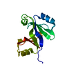
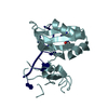
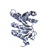
- PDB-4jgl: Crystal structure of a streptavidin-like protein (BACEGG_01519) f... -
+
Open data
ID or keywords:
Loading...
-
Basic information
Entry
Database: PDB / ID: 4jgl
Title
Crystal structure of a streptavidin-like protein (BACEGG_01519) from Bacteroides eggerthii DSM 20697 at 1.25 A resolution
Components
hypothetical protein
Keywords
Structural Genomics / Unknown Function / an orphan / streptavidin-like fold with two extra alpha helices / Joint Center for Structural Genomics / JCSG / Protein Structure Initiative / PSI-BIOLOGY
Mass: 18.015 Da / Num. of mol.: 233 / Source method: isolated from a natural source / Formula: H2O
Has protein modification
Y
Sequence details
THIS CONSTRUCT WAS EXPRESSED WITH A PURIFICATION TAG MGSDKIHHHHHHENLYFQG. THE TAG WAS REMOVED WITH ...THIS CONSTRUCT WAS EXPRESSED WITH A PURIFICATION TAG MGSDKIHHHHHHENLYFQG. THE TAG WAS REMOVED WITH TEV PROTEASE LEAVING ONLY A GLYCINE (0) FOLLOWED BY RESIDUES 24-191 OF THE TARGET SEQUENCE.
-
Experimental details
-
Experiment
Experiment
Method: X-RAY DIFFRACTION / Number of used crystals: 1
-
Sample preparation
Crystal
Density Matthews: 2.39 Å3/Da / Density % sol: 48.59 %
Crystal grow
Temperature: 277 K / Method: vapor diffusion, sitting drop Details: 0.2M calcium acetate, 20.0% polyethylene glycol 3350, NANODROP, VAPOR DIFFUSION, SITTING DROP, temperature 277K
Resolution: 1.25→28.331 Å / Num. all: 49718 / Num. obs: 49718 / % possible obs: 100 % / Redundancy: 11 % / Rsym value: 0.097 / Net I/σ(I): 12.6
Reflection shell
Rmerge(I) obs: 0.014 / Diffraction-ID: 1
Resolution (Å)
Redundancy (%)
Mean I/σ(I) obs
Num. measured all
Num. unique all
Rsym value
% possible all
1.25-1.28
10.9
0.5
39805
3645
1.371
100
1.28-1.32
11
0.7
39307
3584
1.081
100
1.32-1.36
11
0.8
37919
3457
0.914
100
1.36-1.4
11
1
36901
3358
0.752
100
1.4-1.44
11
1.2
36161
3293
0.599
100
1.44-1.49
11
1.6
34528
3131
0.442
100
1.49-1.55
11
2.2
33728
3055
0.335
100
1.55-1.61
11.1
2.9
32678
2956
0.251
100
1.61-1.69
11
3.7
31066
2814
0.198
100
1.69-1.77
11
4.5
29888
2706
0.161
100
1.77-1.86
11
5.5
28137
2549
0.128
100
1.86-1.98
11
6.5
27025
2455
0.104
100
1.98-2.11
10.9
7
24841
2271
0.095
100
2.11-2.28
10.8
7.9
23338
2158
0.083
100
2.28-2.5
10.6
8
20771
1955
0.079
100
2.5-2.8
10.2
8.4
18133
1782
0.074
100
2.8-3.23
11.2
8.8
17627
1574
0.068
100
3.23-3.95
11.6
11
15502
1340
0.055
100
3.95-5.59
11.5
11.5
12005
1045
0.052
100
5.59-28.331
10.9
9.1
6430
590
0.064
99.1
-
Phasing
Phasing
Method: MAD
-
Processing
Software
Name
Version
Classification
NB
MolProbity
3beta29
modelbuilding
PDB_EXTRACT
3.1
dataextraction
SHELX
phasing
SHARP
phasing
SCALA
3.3.20
datascaling
REFMAC
5.5.0110
refinement
MOSFLM
datareduction
SHELXD
phasing
Refinement
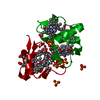
Method to determine structure: MAD / Resolution: 1.25→28.331 Å / Cor.coef. Fo:Fc: 0.98 / Cor.coef. Fo:Fc free: 0.976 / Occupancy max: 1 / Occupancy min: 0.1 / SU B: 1.104 / SU ML: 0.021 / Cross valid method: THROUGHOUT / σ(F): 0 / ESU R: 0.037 / ESU R Free: 0.035 Stereochemistry target values: MAXIMUM LIKELIHOOD WITH PHASES Details: 1. HYDROGENS HAVE BEEN ADDED IN THE RIDING POSITIONS. 2. A MET-INHIBITION PROTOCOL WAS USED FOR SELENOMETHIONINE INCORPORATION DURING PROTEIN EXPRESSION. THE OCCUPANCY OF THE SE ATOMS IN THE ...Details: 1. HYDROGENS HAVE BEEN ADDED IN THE RIDING POSITIONS. 2. A MET-INHIBITION PROTOCOL WAS USED FOR SELENOMETHIONINE INCORPORATION DURING PROTEIN EXPRESSION. THE OCCUPANCY OF THE SE ATOMS IN THE MSE RESIDUES WAS REDUCED TO 0.75 FOR THE REDUCED SCATTERING POWER DUE TO PARTIAL S-MET INCORPORATION. 3. CALCIUM IONS (CA) AND 1,2-ETHANEDIOL (EDO) MOLECULES FROM THE CRYSTALLIZATION/CRYOPROTECTION SOLUTION ARE MODELED. 4. AN UNKNOWN LIAGAND (UNL) HAS BEEN MODELED AT THE SURFACE OF THE PROTEIN BASED ON THE ELECTRON DENSITY. THE DENSITY RESEMBLES NITROBENZENE (NBZ), BENZOIC ACID (BEZ) AND NICOTINIC ACID (NIO). THE EXACT CHEMICAL SPECIES COULD NOT BE DETERMINED WITH THE AVAILABLE DATA.
Rfactor
Num. reflection
% reflection
Selection details
Rfree
0.153
2515
5.1 %
RANDOM
Rwork
0.134
-
-
-
obs
0.135
49663
99.95 %
-
Solvent computation
Ion probe radii: 0.8 Å / Shrinkage radii: 0.8 Å / VDW probe radii: 1.4 Å / Solvent model: BABINET MODEL WITH MASK
In the structure databanks used in Yorodumi, some data are registered as the other names, "COVID-19 virus" and "2019-nCoV". Here are the details of the virus and the list of structure data.
Jan 31, 2019. EMDB accession codes are about to change! (news from PDBe EMDB page)
EMDB accession codes are about to change! (news from PDBe EMDB page)
The allocation of 4 digits for EMDB accession codes will soon come to an end. Whilst these codes will remain in use, new EMDB accession codes will include an additional digit and will expand incrementally as the available range of codes is exhausted. The current 4-digit format prefixed with “EMD-” (i.e. EMD-XXXX) will advance to a 5-digit format (i.e. EMD-XXXXX), and so on. It is currently estimated that the 4-digit codes will be depleted around Spring 2019, at which point the 5-digit format will come into force.
The EM Navigator/Yorodumi systems omit the EMD- prefix.
Related info.:Q: What is EMD? / ID/Accession-code notation in Yorodumi/EM Navigator
Yorodumi is a browser for structure data from EMDB, PDB, SASBDB, etc.
This page is also the successor to EM Navigator detail page, and also detail information page/front-end page for Omokage search.
The word "yorodu" (or yorozu) is an old Japanese word meaning "ten thousand". "mi" (miru) is to see.
Related info.:EMDB / PDB / SASBDB / Comparison of 3 databanks / Yorodumi Search / Aug 31, 2016. New EM Navigator & Yorodumi / Yorodumi Papers / Jmol/JSmol / Function and homology information / Changes in new EM Navigator and Yorodumi
 Movie
Movie Controller

 Yorodumi
 Open data
 Basic information
 Components
 Keywords
 Function and homology information
 Bacteroides eggerthii (bacteria)
 X-RAY DIFFRACTION /
  Authors
 Citation
 Structure visualization
 Downloads & links
 Other downloads
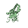


 PDBj
PDBj

 Assembly

 Mass: 40.078 Da / Num. of mol.: 2 / Source method: obtained synthetically / Formula: Ca
 Mass: 62.068 Da / Num. of mol.: 1 / Source method: obtained synthetically / Formula: C2H6O2
 Mass: 18.015 Da / Num. of mol.: 233 / Source method: isolated from a natural source / Formula: H2O
 Sample preparation
 / Beamline: 8.2.2 / Wavelength: 0.918401, 0.979338, 0.979108
 Processing