 Movie
Movie Controller


+ Open data

- Basic information
| Entry | Database: PDB / ID: 4jgg | ||||||
|---|---|---|---|---|---|---|---|
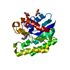
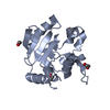
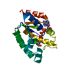
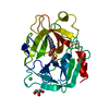
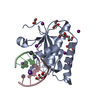
| Title | Crystal Structure of TesA | ||||||
 Components Components | Esterase TesA | ||||||
 Keywords Keywords | HYDROLASE / alpha/beta/alpha / lysophospholipase | ||||||
| Function / homology |  Function and homology information Function and homology informationcarboxylesterase / carboxylesterase activity / phosphatidylcholine lysophospholipase A1 activity / arylesterase activity / extracellular region Similarity search - Function | ||||||
| Biological species |   Pseudomonas aeruginosa (bacteria) Pseudomonas aeruginosa (bacteria) | ||||||
| Method |  X-RAY DIFFRACTION / SYNCHROTRON / MOLECULAR REPLACEMENT / Resolution: 1.9 Å X-RAY DIFFRACTION / SYNCHROTRON / MOLECULAR REPLACEMENT / Resolution: 1.9 Å | ||||||
 Authors Authors | Kovacic, F. / Granzin, J. / Batra-Safferling, R. / Jaeger, K.-E. | ||||||
 Citation Citation | Journal: Plos One / Year: 2013 Title: Structural and Functional Characterisation of TesA - A Novel Lysophospholipase A from Pseudomonas aeruginosa. Authors: Kovacic, F. / Granzin, J. / Wilhelm, S. / Kojic-Prodic, B. / Batra-Safferling, R. / Jaeger, K.E. | ||||||
| History |
|
- Structure visualization
| Structure viewer | Molecule: MolmilJmol/JSmol |
|---|
- Downloads & links
-Download
| PDBx/mmCIF format | 4jgg.cif.gz | 82.9 KB | Display | PDBx/mmCIF format |
|---|---|---|---|---|
| PDB format | pdb4jgg.ent.gz | 61.6 KB | Display | PDB format |
| PDBx/mmJSON format | 4jgg.json.gz | Tree view | PDBx/mmJSON format | |
| Others |  Other downloads Other downloads |
-Validation report
| Arichive directory | https://data.pdbj.org/pub/pdb/validation_reports/jg/4jggftp://data.pdbj.org/pub/pdb/validation_reports/jg/4jgg | HTTPS FTP |
|---|
-Related structure data
| Related structure data | 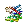 3hp4S S: Starting model for refinement |
|---|---|
| Similar structure data |


-Links
 PDBj
PDBj- Assembly
| Deposited unit | 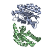
| ||||||||
|---|---|---|---|---|---|---|---|---|---|
| 1 | 
| ||||||||
| 2 | 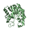
| ||||||||
| Unit cell |
|
-Components
| #1: Protein | Mass: 21882.291 Da / Num. of mol.: 2 Source method: isolated from a genetically manipulated source Source: (gene. exp.) Pseudomonas aeruginosa (bacteria) / Strain: ATCC 15692 / PAO1 / 1C / PRS 101 / LMG 12228 / Gene: PA2856, tesA / Plasmid: pET22b / Production host: #2: Water | ChemComp-HOH / |  Mass: 18.015 Da / Num. of mol.: 276 / Source method: isolated from a natural source / Formula: H2O Mass: 18.015 Da / Num. of mol.: 276 / Source method: isolated from a natural source / Formula: H2O |
|---|
-Experimental details
-Experiment
| Experiment | Method: X-RAY DIFFRACTION / Number of used crystals: 1 |
|---|
- Sample preparation
| Crystal | Density Matthews: 2.2 Å3/Da / Density % sol: 43.97 % |
|---|---|
| Crystal grow | Temperature: 292 K / Method: vapor diffusion, sitting drop / pH: 4.5 Details: 20% PEG 3350, 50mM sodium-citrate, pH 4.5, VAPOR DIFFUSION, SITTING DROP, temperature 292K |
-Data collection
| Diffraction | Mean temperature: 100 K |
|---|---|
| Diffraction source | Source: SYNCHROTRON / Site: ESRF  / Beamline: ID14-1 / Wavelength: 0.9334 Å / Beamline: ID14-1 / Wavelength: 0.9334 Å |
| Detector | Type: ADSC QUANTUM 210 / Detector: CCD / Date: Nov 4, 2009 |
| Radiation | Monochromator: Diamond (111), Ge(220) / Protocol: SINGLE WAVELENGTH / Monochromatic (M) / Laue (L): M / Scattering type: x-ray |
| Radiation wavelength | Wavelength: 0.9334 Å / Relative weight: 1 |
| Reflection | Resolution: 1.9→45.49 Å / Num. all: 29767 / Num. obs: 29422 / % possible obs: 98.9 % / Observed criterion σ(F): 0 / Observed criterion σ(I): 0 / Redundancy: 3.2 % / Biso Wilson estimate: 13.45 Å2 / Rmerge(I) obs: 0.064 / Rsym value: 0.064 / Net I/σ(I): 13 |
| Reflection shell | Resolution: 1.9→1.93 Å / Redundancy: 3.2 % / Rmerge(I) obs: 0.242 / Mean I/σ(I) obs: 4.7 / Num. unique all: 1485 / Rsym value: 0.242 / % possible all: 98.9 |
- Processing
| Software |
| |||||||||||||||||||||||||||||||||||||||||||||||||||||||||||||||||||||||||||||
|---|---|---|---|---|---|---|---|---|---|---|---|---|---|---|---|---|---|---|---|---|---|---|---|---|---|---|---|---|---|---|---|---|---|---|---|---|---|---|---|---|---|---|---|---|---|---|---|---|---|---|---|---|---|---|---|---|---|---|---|---|---|---|---|---|---|---|---|---|---|---|---|---|---|---|---|---|---|---|
| Refinement | Method to determine structure: MOLECULAR REPLACEMENT Starting model: PDB ENTRY 3HP4 Resolution: 1.9→40.625 Å / SU ML: 0.26 / Cross valid method: THROUGHOUT / σ(F): 0 / Phase error: 28.3 / Stereochemistry target values: Engh & Huber
| |||||||||||||||||||||||||||||||||||||||||||||||||||||||||||||||||||||||||||||
| Displacement parameters |
| |||||||||||||||||||||||||||||||||||||||||||||||||||||||||||||||||||||||||||||
| Refinement step | Cycle: LAST / Resolution: 1.9→40.625 Å
| |||||||||||||||||||||||||||||||||||||||||||||||||||||||||||||||||||||||||||||
| Refine LS restraints |
| |||||||||||||||||||||||||||||||||||||||||||||||||||||||||||||||||||||||||||||
| LS refinement shell |
|
