 Movie
Movie Controller

[English] 日本語
 Yorodumi
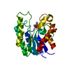
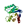
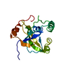
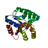
- PDB-3h5j: LeuD_1-168 small subunit of isopropylmalate isomerase (Rv2987c) f... -
+ Open data

- Basic information
| Entry | Database: PDB / ID: 3h5j | ||||||
|---|---|---|---|---|---|---|---|
| Title | LeuD_1-168 small subunit of isopropylmalate isomerase (Rv2987c) from mycobacterium tuberculosis | ||||||
 Components Components | 3-isopropylmalate dehydratase small subunit | ||||||
 Keywords Keywords | LYASE / Leucine biosynthesis / isopropylmalate isomerase / LeuD / M.tuberculosis / Amino-acid biosynthesis / Branched-chain amino acid biosynthesis | ||||||
| Function / homology |  Function and homology information Function and homology information3-isopropylmalate dehydratase complex / 3-isopropylmalate dehydratase / 3-isopropylmalate dehydratase activity / L-leucine biosynthetic process / plasma membrane Similarity search - Function | ||||||
| Biological species |   Mycobacterium tuberculosis (bacteria) Mycobacterium tuberculosis (bacteria) | ||||||
| Method |  X-RAY DIFFRACTION / SYNCHROTRON / MOLECULAR REPLACEMENT / Resolution: 1.2 Å X-RAY DIFFRACTION / SYNCHROTRON / MOLECULAR REPLACEMENT / Resolution: 1.2 Å | ||||||
 Authors Authors | Manikandan, K. / Geerlof, A. / Weiss, M.S. | ||||||
 Citation Citation | Journal: Proteins / Year: 2011 Title: Structural studies on the enzyme complex isopropylmalate isomerase (LeuCD) from Mycobacterium tuberculosis Authors: Manikandan, K. / Geerlof, A. / Zozulya, A.V. / Svergun, D.I. / Weiss, M.S. #1: Journal: Acta Crystallogr.,Sect.F / Year: 2009 Title: Cloning, expression, purification, crystallization and preliminary X-ray diffraction analysis of the small subunit of isopropylmalate isomerase (Rv2987c) from Mycobacterium tuberculosis Authors: Manikandan, K. / Geerlof, A. / Schuldt, L. / Mueller-Dieckmann, C. / Weiss, M.S. | ||||||
| History |
|
- Structure visualization
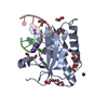
| Structure viewer | Molecule: MolmilJmol/JSmol |
|---|
- Downloads & links
-Download
| PDBx/mmCIF format | 3h5j.cif.gz | 167.5 KB | Display | PDBx/mmCIF format |
|---|---|---|---|---|
| PDB format | pdb3h5j.ent.gz | 131.9 KB | Display | PDB format |
| PDBx/mmJSON format | 3h5j.json.gz | Tree view | PDBx/mmJSON format | |
| Others |  Other downloads Other downloads |
-Validation report
| Arichive directory | https://data.pdbj.org/pub/pdb/validation_reports/h5/3h5jftp://data.pdbj.org/pub/pdb/validation_reports/h5/3h5j | HTTPS FTP |
|---|
-Related structure data
| Related structure data |  3h5eSC  3h5hC S: Starting model for refinement C: citing same article ( |
|---|---|
| Similar structure data |


-Links
 PDBj
PDBj

- Assembly
| Deposited unit | 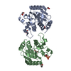
| ||||||||
|---|---|---|---|---|---|---|---|---|---|
| 1 | 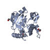
| ||||||||
| 2 | 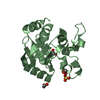
| ||||||||
| Unit cell |
| ||||||||
| Details | AS THE STRUCTURE REPRESENTS ONLY A PART OF A SUBUNIT OF AN ENZYME COMPLEX, THE ASSEMBLIES DESCRIBED IN REMARK350 IS NOT RELEVANT TO THE REAL BIOLOGICAL ASSEMBLIES |
-Components
| #1: Protein | Mass: 18644.068 Da / Num. of mol.: 2 / Fragment: LeuD, residues 1-168 Source method: isolated from a genetically manipulated source Source: (gene. exp.) Mycobacterium tuberculosis (bacteria) / Strain: H37Rv / Gene: Rv2987c / Plasmid: pETM11 / Production host: References: UniProt: P65277, UniProt: P9WK95*PLUS, 3-isopropylmalate dehydratase #2: Chemical | ChemComp-EDO /   Mass: 62.068 Da / Num. of mol.: 7 / Source method: obtained synthetically / Formula: C2H6O2 Mass: 62.068 Da / Num. of mol.: 7 / Source method: obtained synthetically / Formula: C2H6O2#3: Chemical | ChemComp-SO4 / |   Mass: 96.063 Da / Num. of mol.: 1 / Source method: obtained synthetically / Formula: SO4 Mass: 96.063 Da / Num. of mol.: 1 / Source method: obtained synthetically / Formula: SO4#4: Water | ChemComp-HOH / |  Mass: 18.015 Da / Num. of mol.: 413 / Source method: isolated from a natural source / Formula: H2O Mass: 18.015 Da / Num. of mol.: 413 / Source method: isolated from a natural source / Formula: H2OSequence details | THE SER 1A WAS INTRODUCED | |
|---|
-Experimental details
-Experiment
| Experiment | Method: X-RAY DIFFRACTION / Number of used crystals: 1 |
|---|
- Sample preparation
| Crystal | Density Matthews: 1.98 Å3/Da / Density % sol: 37.95 % |
|---|---|
| Crystal grow | Temperature: 293 K / Method: vapor diffusion / pH: 8.5 Details: 0.2M lithium sulfate, 0.1M Tris-HCl pH 8.5, 20%(w/v) PEG 3350, VAPOR DIFFUSION, temperature 293K |
-Data collection
| Diffraction | Mean temperature: 100 K |
|---|---|
| Diffraction source | Source: SYNCHROTRON / Site: EMBL/DESY, HAMBURG  / Beamline: X12 / Wavelength: 1 Å / Beamline: X12 / Wavelength: 1 Å |
| Detector | Type: MARMOSAIC 225 mm CCD / Detector: CCD / Date: Oct 22, 2008 |
| Radiation | Monochromator: Double crystal Si(111) / Protocol: SINGLE WAVELENGTH / Monochromatic (M) / Laue (L): M / Scattering type: x-ray |
| Radiation wavelength | Wavelength: 1 Å / Relative weight: 1 |
| Reflection | Resolution: 1.2→99 Å / Num. all: 90248 / Num. obs: 90248 / % possible obs: 99.5 % / Observed criterion σ(F): 2 / Observed criterion σ(I): 2 / Redundancy: 4 % / Rmerge(I) obs: 0.065 / Net I/σ(I): 20.7 |
| Reflection shell | Resolution: 1.2→1.22 Å / Redundancy: 3.4 % / Rmerge(I) obs: 0.296 / Mean I/σ(I) obs: 3.8 / % possible all: 91.9 |
- Processing
| Software |
| |||||||||||||||||||||||||||||||||
|---|---|---|---|---|---|---|---|---|---|---|---|---|---|---|---|---|---|---|---|---|---|---|---|---|---|---|---|---|---|---|---|---|---|---|
| Refinement | Method to determine structure: MOLECULAR REPLACEMENT Starting model: PDB ENTRY 3H5E Resolution: 1.2→13.42 Å / Num. parameters: 28335 / Num. restraintsaints: 35073 / Cross valid method: FREE R / σ(F): 0 / Stereochemistry target values: Engh & Huber
| |||||||||||||||||||||||||||||||||
| Refine analyze | Num. disordered residues: 24 / Occupancy sum hydrogen: 2546.15 / Occupancy sum non hydrogen: 3024.55 | |||||||||||||||||||||||||||||||||
| Refinement step | Cycle: LAST / Resolution: 1.2→13.42 Å
| |||||||||||||||||||||||||||||||||
| Refine LS restraints |
|
