 Movie
Movie Controller

[English] 日本語
 Yorodumi
- PDB-4jg7: Structure of RSK2 CTD bound to 3-(3-(1H-pyrrolo[2,3-b]pyridine-3-... -
+ Open data

- Basic information
| Entry | Database: PDB / ID: 4jg7 | ||||||
|---|---|---|---|---|---|---|---|
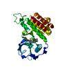
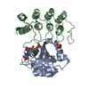
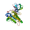
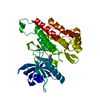
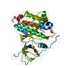
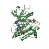
| Title | Structure of RSK2 CTD bound to 3-(3-(1H-pyrrolo[2,3-b]pyridine-3-carbonyl)phenyl)-2-cyanoacrylamide | ||||||
 Components Components | Ribosomal protein S6 kinase alpha-3 | ||||||
 Keywords Keywords | TRANSFERASE/TRANSFERASE INHIBITOR / Protein Kinase / Phosphorylation / Covalent Inhibitor / TRANSFERASE-TRANSFERASE INHIBITOR complex | ||||||
| Function / homology |  Function and homology information Function and homology informationregulation of translation in response to stress / CREB1 phosphorylation through NMDA receptor-mediated activation of RAS signaling / ribosomal protein S6 kinase activity / CREB phosphorylation / Gastrin-CREB signalling pathway via PKC and MAPK / RSK activation / toll-like receptor signaling pathway / ERK/MAPK targets / TORC1 signaling / Recycling pathway of L1 ...regulation of translation in response to stress / CREB1 phosphorylation through NMDA receptor-mediated activation of RAS signaling / ribosomal protein S6 kinase activity / CREB phosphorylation / Gastrin-CREB signalling pathway via PKC and MAPK / RSK activation / toll-like receptor signaling pathway / ERK/MAPK targets / TORC1 signaling / Recycling pathway of L1 / skeletal system development / central nervous system development / positive regulation of cell differentiation / positive regulation of cell growth / Senescence-Associated Secretory Phenotype (SASP) / response to lipopolysaccharide / chemical synaptic transmission / protein kinase activity / non-specific serine/threonine protein kinase / protein serine kinase activity / protein serine/threonine kinase activity / regulation of DNA-templated transcription / synapse / nucleolus / negative regulation of apoptotic process / protein kinase binding / magnesium ion binding / signal transduction / positive regulation of transcription by RNA polymerase II / nucleoplasm / ATP binding / cytosol / cytoplasm Similarity search - Function | ||||||
| Biological species |  Homo sapiens (human) Homo sapiens (human) | ||||||
| Method |  X-RAY DIFFRACTION / MOLECULAR REPLACEMENT / Resolution: 3.0002 Å X-RAY DIFFRACTION / MOLECULAR REPLACEMENT / Resolution: 3.0002 Å | ||||||
 Authors Authors | Miller, R.M. / Paavilainen, V.O. / Krishnan, S. / Serafimova, I.M. / Taunton, J. | ||||||
 Citation Citation | Journal: J.Am.Chem.Soc. / Year: 2013 Title: Electrophilic fragment-based design of reversible covalent kinase inhibitors. Authors: Miller, R.M. / Paavilainen, V.O. / Krishnan, S. / Serafimova, I.M. / Taunton, J. | ||||||
| History |
|
- Structure visualization
| Structure viewer | Molecule: MolmilJmol/JSmol |
|---|
- Downloads & links
-Download
| PDBx/mmCIF format | 4jg7.cif.gz | 122.8 KB | Display | PDBx/mmCIF format |
|---|---|---|---|---|
| PDB format | pdb4jg7.ent.gz | 94.5 KB | Display | PDB format |
| PDBx/mmJSON format | 4jg7.json.gz | Tree view | PDBx/mmJSON format | |
| Others |  Other downloads Other downloads |
-Validation report
| Arichive directory | https://data.pdbj.org/pub/pdb/validation_reports/jg/4jg7ftp://data.pdbj.org/pub/pdb/validation_reports/jg/4jg7 | HTTPS FTP |
|---|
-Related structure data
| Related structure data | 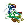 4jg6C  4jg8C  2qr8S S: Starting model for refinement C: citing same article ( |
|---|---|
| Similar structure data |


-Links
 PDBj
PDBj


- Assembly
| Deposited unit | 
| ||||||||
|---|---|---|---|---|---|---|---|---|---|
| 1 |
| ||||||||
| Unit cell |
| ||||||||
| Details | biological unit is the same as asym. |
-Components
| #1: Protein | Mass: 40067.551 Da / Num. of mol.: 1 Source method: isolated from a genetically manipulated source Source: (gene. exp.) Homo sapiens (human) / Gene: RPS6KA3, ISPK1, MAPKAPK1B, RSK2 / Production host:  References: UniProt: P51812, non-specific serine/threonine protein kinase |
|---|---|
| #2: Chemical | ChemComp-1LC / (  Mass: 318.329 Da / Num. of mol.: 1 / Source method: obtained synthetically / Formula: C18H14N4O2 Mass: 318.329 Da / Num. of mol.: 1 / Source method: obtained synthetically / Formula: C18H14N4O2 |
| #3: Chemical | ChemComp-NA /   Mass: 22.990 Da / Num. of mol.: 1 / Source method: obtained synthetically / Formula: Na Mass: 22.990 Da / Num. of mol.: 1 / Source method: obtained synthetically / Formula: Na |
| #4: Water | ChemComp-HOH /  Mass: 18.015 Da / Num. of mol.: 22 / Source method: isolated from a natural source / Formula: H2O Mass: 18.015 Da / Num. of mol.: 22 / Source method: isolated from a natural source / Formula: H2O |
| Has protein modification | Y |
-Experimental details
-Experiment
| Experiment | Method: X-RAY DIFFRACTION / Number of used crystals: 1 |
|---|
- Sample preparation
| Crystal | Density Matthews: 2.07 Å3/Da / Density % sol: 40.58 % |
|---|---|
| Crystal grow | Temperature: 298 K / Method: vapor diffusion, hanging drop / pH: 7 Details: 100 mM HEPES, 50 mM Ammonium Sulfate, 7.5% PEG3350, pH 7.0, VAPOR DIFFUSION, HANGING DROP, temperature 298K |
-Data collection
| Diffraction | Mean temperature: 100 K |
|---|---|
| Diffraction source | Source: ROTATING ANODE / Type: RIGAKU RU200 / Wavelength: 1.5418 Å |
| Detector | Type: RIGAKU RAXIS IV / Detector: IMAGE PLATE / Date: Aug 4, 2009 |
| Radiation | Monochromator: Flat Graphite Crystal / Protocol: SINGLE WAVELENGTH / Monochromatic (M) / Laue (L): M / Scattering type: x-ray |
| Radiation wavelength | Wavelength: 1.5418 Å / Relative weight: 1 |
| Reflection | Resolution: 3→47.6 Å / Num. all: 7446 / Num. obs: 7417 / % possible obs: 99.3 % / Observed criterion σ(F): 3 / Observed criterion σ(I): 3 |
| Reflection shell | Resolution: 3→3.18 Å / % possible all: 99.3 |
- Processing
| Software |
| ||||||||||||||||||||||||||||||||||||||||||
|---|---|---|---|---|---|---|---|---|---|---|---|---|---|---|---|---|---|---|---|---|---|---|---|---|---|---|---|---|---|---|---|---|---|---|---|---|---|---|---|---|---|---|---|
| Refinement | Method to determine structure: MOLECULAR REPLACEMENT Starting model: PDB ENTRY 2QR8 Resolution: 3.0002→47.038 Å / Occupancy max: 1 / Occupancy min: 0.31 / SU ML: 0.91 / σ(F): 0 / Phase error: 26.97 / Stereochemistry target values: ML
| ||||||||||||||||||||||||||||||||||||||||||
| Solvent computation | Shrinkage radii: 0.77 Å / VDW probe radii: 0.9 Å / Solvent model: FLAT BULK SOLVENT MODEL / Bsol: 10.943 Å2 / ksol: 0.358 e/Å3 | ||||||||||||||||||||||||||||||||||||||||||
| Displacement parameters |
| ||||||||||||||||||||||||||||||||||||||||||
| Refinement step | Cycle: LAST / Resolution: 3.0002→47.038 Å
| ||||||||||||||||||||||||||||||||||||||||||
| Refine LS restraints |
| ||||||||||||||||||||||||||||||||||||||||||
| LS refinement shell |
|
