 Movie
Movie Controller

[English] 日本語
 Yorodumi
- PDB-4jav: Structural basis of a rationally rewired protein-protein interfac... -
+ Open data

- Basic information
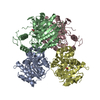
| Entry | Database: PDB / ID: 4jav | ||||||
|---|---|---|---|---|---|---|---|
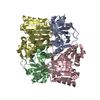
| Title | Structural basis of a rationally rewired protein-protein interface (HK853wt and RR468mutant V13P, L14I, I17M and N21V) | ||||||
 Components Components |
| ||||||
 Keywords Keywords | TRANSFERASE/SIGNALING PROTEIN / Bergerat fold / Four helix bundle / alpha/beta fold / Signal transduction / Histidine kinase / autophosphorylation / phosphotransferase / dephosphorylation / TRANSFERASE-SIGNALING PROTEIN complex | ||||||
| Function / homology |  Function and homology information Function and homology informationhistidine phosphotransfer kinase activity / phosphorelay response regulator activity / phosphorelay sensor kinase activity / histidine kinase / phosphorelay signal transduction system / DNA-templated transcription / DNA binding / ATP binding / metal ion binding / plasma membrane Similarity search - Function | ||||||
| Biological species |   Thermotoga maritima (bacteria) Thermotoga maritima (bacteria) | ||||||
| Method |  X-RAY DIFFRACTION / SYNCHROTRON / MOLECULAR REPLACEMENT / Resolution: 3.1 Å X-RAY DIFFRACTION / SYNCHROTRON / MOLECULAR REPLACEMENT / Resolution: 3.1 Å | ||||||
 Authors Authors | Podgornaia, A.I. / Casino, P. / Marina, A. / Laub, M.T. | ||||||
 Citation Citation | Journal: Structure / Year: 2013 Title: Structural basis of a rationally rewired protein-protein interface critical to bacterial signaling Authors: Podgornaia, A.I. / Casino, P. / Marina, A. / Laub, M.T. | ||||||
| History |
|
- Structure visualization
| Structure viewer | Molecule: MolmilJmol/JSmol |
|---|
- Downloads & links
-Download
| PDBx/mmCIF format | 4jav.cif.gz | 160.5 KB | Display | PDBx/mmCIF format |
|---|---|---|---|---|
| PDB format | pdb4jav.ent.gz | 126.4 KB | Display | PDB format |
| PDBx/mmJSON format | 4jav.json.gz | Tree view | PDBx/mmJSON format | |
| Others |  Other downloads Other downloads |
-Validation report
| Arichive directory | https://data.pdbj.org/pub/pdb/validation_reports/ja/4javftp://data.pdbj.org/pub/pdb/validation_reports/ja/4jav | HTTPS FTP |
|---|
-Related structure data
| Related structure data | 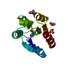 4ja2C  4jasC  4jauC  2c2aS 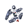 3gl9S C: citing same article ( S: Starting model for refinement |
|---|---|
| Similar structure data |

-Links
 PDBj
PDBj


- Assembly
| Deposited unit | 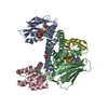
| ||||||||
|---|---|---|---|---|---|---|---|---|---|
| 1 |
| ||||||||
| Unit cell |
|
-Components
-Protein , 2 types, 4 molecules ABCD
| #1: Protein | Mass: 29378.281 Da / Num. of mol.: 2 / Fragment: HK853 cytoplasmic region, UNP residues 232-489 Source method: isolated from a genetically manipulated source Source: (gene. exp.) Thermotoga maritima (bacteria) / Strain: MSB8 / Gene: TM_0853 / Plasmid: pET24 / Production host: #2: Protein | Mass: 13914.305 Da / Num. of mol.: 2 / Mutation: V13P, L14I, I17M, N21V Source method: isolated from a genetically manipulated source Source: (gene. exp.) Thermotoga maritima (bacteria) / Strain: MSB8 / Gene: TM_0468 / Plasmid: pET22 / Production host: |
|---|
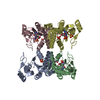
-Non-polymers , 5 types, 25 molecules 


| #3: Chemical | ChemComp-SO4 /  Mass: 96.063 Da / Num. of mol.: 5 / Source method: obtained synthetically / Formula: SO4 Mass: 96.063 Da / Num. of mol.: 5 / Source method: obtained synthetically / Formula: SO4#4: Chemical | ChemComp-MG /  Mass: 24.305 Da / Num. of mol.: 4 / Source method: obtained synthetically / Formula: Mg Mass: 24.305 Da / Num. of mol.: 4 / Source method: obtained synthetically / Formula: Mg#5: Chemical |  Mass: 427.201 Da / Num. of mol.: 2 / Source method: obtained synthetically / Formula: C10H15N5O10P2 / Comment: ADP, energy-carrying molecule*YM Mass: 427.201 Da / Num. of mol.: 2 / Source method: obtained synthetically / Formula: C10H15N5O10P2 / Comment: ADP, energy-carrying molecule*YM#6: Chemical | ChemComp-CL / |  Mass: 35.453 Da / Num. of mol.: 1 / Source method: obtained synthetically / Formula: Cl Mass: 35.453 Da / Num. of mol.: 1 / Source method: obtained synthetically / Formula: Cl#7: Water | ChemComp-HOH / | Mass: 18.015 Da / Num. of mol.: 13 / Source method: isolated from a natural source / Formula: H2O |
|---|
-Details
| Has protein modification | Y |
|---|
-Experimental details
-Experiment
| Experiment | Method: X-RAY DIFFRACTION / Number of used crystals: 1 |
|---|
- Sample preparation
| Crystal | Density Matthews: 3.45 Å3/Da / Density % sol: 64.3 % |
|---|---|
| Crystal grow | Temperature: 294 K / Method: vapor diffusion, sitting drop / pH: 5.5 Details: 2.2M ammonium sulfate, Bis-Tris, pH 5.5, VAPOR DIFFUSION, SITTING DROP, temperature 294K |
-Data collection
| Diffraction | Mean temperature: 100 K |
|---|---|
| Diffraction source | Source: SYNCHROTRON / Site: ESRF  / Beamline: ID23-2 / Wavelength: 0.8726 Å / Beamline: ID23-2 / Wavelength: 0.8726 Å |
| Detector | Type: MARMOSAIC 225 mm CCD / Detector: CCD / Date: Sep 29, 2011 Details: Focusing mirrors: one pair of (300x40x15) mm3 long Pt coated Si mirror, 260mm usable, in a Kirkpatrick-Baez geometry |
| Radiation | Monochromator: horizontally diffracting monochromator / Protocol: SINGLE WAVELENGTH / Monochromatic (M) / Laue (L): M / Scattering type: x-ray |
| Radiation wavelength | Wavelength: 0.8726 Å / Relative weight: 1 |
| Reflection | Resolution: 3.1→46.3 Å / Num. obs: 22038 / % possible obs: 99.9 % / Observed criterion σ(F): 1.9 / Observed criterion σ(I): 2 / Rmerge(I) obs: 0.071 |
| Reflection shell | Resolution: 3.1→3.27 Å / Redundancy: 4.9 % / Rmerge(I) obs: 0.401 / Mean I/σ(I) obs: 17.9 / Num. unique all: 22038 / Rsym value: 0.401 / % possible all: 99.9 |
- Processing
| Software |
| ||||||||||||||||||||||||||||||||||||||||||||||||||||||||||||||||||||||||||||||||||||||||||||||||||||||||||||||||||||||||||||||||||||||||||||||||||||||||||||||||||||||||||
|---|---|---|---|---|---|---|---|---|---|---|---|---|---|---|---|---|---|---|---|---|---|---|---|---|---|---|---|---|---|---|---|---|---|---|---|---|---|---|---|---|---|---|---|---|---|---|---|---|---|---|---|---|---|---|---|---|---|---|---|---|---|---|---|---|---|---|---|---|---|---|---|---|---|---|---|---|---|---|---|---|---|---|---|---|---|---|---|---|---|---|---|---|---|---|---|---|---|---|---|---|---|---|---|---|---|---|---|---|---|---|---|---|---|---|---|---|---|---|---|---|---|---|---|---|---|---|---|---|---|---|---|---|---|---|---|---|---|---|---|---|---|---|---|---|---|---|---|---|---|---|---|---|---|---|---|---|---|---|---|---|---|---|---|---|---|---|---|---|---|---|---|
| Refinement | Method to determine structure: MOLECULAR REPLACEMENT Starting model: 2C2A, 3GL9 Resolution: 3.1→44.55 Å / Cor.coef. Fo:Fc: 0.938 / Cor.coef. Fo:Fc free: 0.909 / SU B: 19.224 / SU ML: 0.332 / Cross valid method: THROUGHOUT / ESU R Free: 0.426 / Stereochemistry target values: MAXIMUM LIKELIHOOD / Details: HYDROGENS HAVE BEEN USED IF PRESENT IN THE INPUT
| ||||||||||||||||||||||||||||||||||||||||||||||||||||||||||||||||||||||||||||||||||||||||||||||||||||||||||||||||||||||||||||||||||||||||||||||||||||||||||||||||||||||||||
| Solvent computation | Ion probe radii: 0.8 Å / Shrinkage radii: 0.8 Å / VDW probe radii: 1.2 Å / Solvent model: MASK | ||||||||||||||||||||||||||||||||||||||||||||||||||||||||||||||||||||||||||||||||||||||||||||||||||||||||||||||||||||||||||||||||||||||||||||||||||||||||||||||||||||||||||
| Displacement parameters | Biso mean: 71.969 Å2
| ||||||||||||||||||||||||||||||||||||||||||||||||||||||||||||||||||||||||||||||||||||||||||||||||||||||||||||||||||||||||||||||||||||||||||||||||||||||||||||||||||||||||||
| Refinement step | Cycle: LAST / Resolution: 3.1→44.55 Å
| ||||||||||||||||||||||||||||||||||||||||||||||||||||||||||||||||||||||||||||||||||||||||||||||||||||||||||||||||||||||||||||||||||||||||||||||||||||||||||||||||||||||||||
| Refine LS restraints |
| ||||||||||||||||||||||||||||||||||||||||||||||||||||||||||||||||||||||||||||||||||||||||||||||||||||||||||||||||||||||||||||||||||||||||||||||||||||||||||||||||||||||||||
| LS refinement shell | Resolution: 3.1→3.18 Å / Total num. of bins used: 20
|
