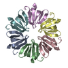
 Movie
Movie Controller

[English] 日本語
 Yorodumi
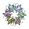
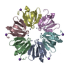
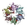
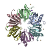
- PDB-4j6w: Crystal structure of HFQ from Pseudomonas aeruginosa in complex w... -
+ Open data

- Basic information
| Entry | Database: PDB / ID: 4j6w | |||||||||
|---|---|---|---|---|---|---|---|---|---|---|
| Title | Crystal structure of HFQ from Pseudomonas aeruginosa in complex with CTP | |||||||||
 Components Components | Protein hfq | |||||||||
 Keywords Keywords | RNA BINDING PROTEIN / LSM / RNA chaperone / sRNA / mRNA | |||||||||
| Function / homology |  Function and homology information Function and homology informationregulation of translation, ncRNA-mediated / regulation of carbon utilization / regulation of RNA stability / quorum sensing / regulation of translation / regulation of DNA-templated transcription / RNA binding / cytosol Similarity search - Function | |||||||||
| Biological species |   Pseudomonas aeruginosa (bacteria) Pseudomonas aeruginosa (bacteria) | |||||||||
| Method |  X-RAY DIFFRACTION / SYNCHROTRON / MOLECULAR REPLACEMENT / Resolution: 1.8 Å X-RAY DIFFRACTION / SYNCHROTRON / MOLECULAR REPLACEMENT / Resolution: 1.8 Å | |||||||||
 Authors Authors | Nikulin, A.D. / Murina, V. / Lekontseva, N. | |||||||||
 Citation Citation | Journal: Acta Crystallogr.,Sect.D / Year: 2013 Title: Hfq binds ribonucleotides in three different RNA-binding sites. Authors: Murina, V. / Lekontseva, N. / Nikulin, A. | |||||||||
| History |
|
- Structure visualization
| Structure viewer | Molecule: MolmilJmol/JSmol |
|---|
- Downloads & links
-Download
| PDBx/mmCIF format | 4j6w.cif.gz | 113.4 KB | Display | PDBx/mmCIF format |
|---|---|---|---|---|
| PDB format | pdb4j6w.ent.gz | 88.8 KB | Display | PDB format |
| PDBx/mmJSON format | 4j6w.json.gz | Tree view | PDBx/mmJSON format | |
| Others |  Other downloads Other downloads |
-Validation report
| Arichive directory | https://data.pdbj.org/pub/pdb/validation_reports/j6/4j6wftp://data.pdbj.org/pub/pdb/validation_reports/j6/4j6w | HTTPS FTP |
|---|
-Related structure data
| Related structure data |  3quiC 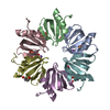 4j5yC 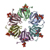 4j6xC 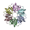 4j6yC 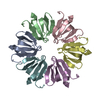 1u1tS S: Starting model for refinement C: citing same article ( |
|---|---|
| Similar structure data |


-Links
 PDBj
PDBj


- Assembly
| Deposited unit | 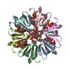
| ||||||||
|---|---|---|---|---|---|---|---|---|---|
| 1 |
| ||||||||
| Unit cell |
|
-Components
-Protein , 1 types, 6 molecules ABCDEF
| #1: Protein | Mass: 9114.487 Da / Num. of mol.: 6 Source method: isolated from a genetically manipulated source Source: (gene. exp.) Pseudomonas aeruginosa (bacteria) / Strain: ATCC 15692 / PAO1 / 1C / PRS 101 / LMG 12228 / Gene: hfq, PA4944 / Plasmid: PET22B / Production host: |
|---|
-Non-polymers , 8 types, 323 molecules 


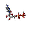

| #2: Chemical | ChemComp-CTP /  Mass: 483.156 Da / Num. of mol.: 6 / Source method: obtained synthetically / Formula: C9H16N3O14P3 Mass: 483.156 Da / Num. of mol.: 6 / Source method: obtained synthetically / Formula: C9H16N3O14P3#3: Chemical | ChemComp-C5P /  Mass: 323.197 Da / Num. of mol.: 4 / Source method: obtained synthetically / Formula: C9H14N3O8P Mass: 323.197 Da / Num. of mol.: 4 / Source method: obtained synthetically / Formula: C9H14N3O8P#4: Chemical | ChemComp-MG /  Mass: 24.305 Da / Num. of mol.: 8 / Source method: obtained synthetically / Formula: Mg Mass: 24.305 Da / Num. of mol.: 8 / Source method: obtained synthetically / Formula: Mg#5: Chemical |  Mass: 22.990 Da / Num. of mol.: 3 / Source method: obtained synthetically / Formula: Na Mass: 22.990 Da / Num. of mol.: 3 / Source method: obtained synthetically / Formula: Na#6: Chemical | ChemComp-P6G / |  Mass: 282.331 Da / Num. of mol.: 1 / Source method: obtained synthetically / Formula: C12H26O7 / Comment: precipitant*YM Mass: 282.331 Da / Num. of mol.: 1 / Source method: obtained synthetically / Formula: C12H26O7 / Comment: precipitant*YM#7: Chemical |  Mass: 35.453 Da / Num. of mol.: 2 / Source method: obtained synthetically / Formula: Cl Mass: 35.453 Da / Num. of mol.: 2 / Source method: obtained synthetically / Formula: Cl#8: Chemical | ChemComp-CDP / |  Mass: 403.176 Da / Num. of mol.: 1 / Source method: obtained synthetically / Formula: C9H15N3O11P2 Mass: 403.176 Da / Num. of mol.: 1 / Source method: obtained synthetically / Formula: C9H15N3O11P2#9: Water | ChemComp-HOH / | Mass: 18.015 Da / Num. of mol.: 298 / Source method: isolated from a natural source / Formula: H2O |
|---|
-Experimental details
-Experiment
| Experiment | Method: X-RAY DIFFRACTION / Number of used crystals: 1 |
|---|
- Sample preparation
| Crystal | Density Matthews: 2.29 Å3/Da / Density % sol: 46.27 % |
|---|---|
| Crystal grow | Temperature: 295 K / Method: vapor diffusion, hanging drop / pH: 8 Details: 200 mm ammonium sulphate, 200 mm NaCl, 50 mm TRIS-HCl, pH 8.0, VAPOR DIFFUSION, HANGING DROP, temperature 295K |
-Data collection
| Diffraction | Mean temperature: 100 K |
|---|---|
| Diffraction source | Source: SYNCHROTRON / Site: BESSY  / Beamline: 14.1 / Wavelength: 0.91841 Å / Beamline: 14.1 / Wavelength: 0.91841 Å |
| Detector | Type: MARMOSAIC 225 mm CCD / Detector: CCD / Date: May 10, 2012 |
| Radiation | Monochromator: SI-111 CRYSTAL / Protocol: SINGLE WAVELENGTH / Monochromatic (M) / Laue (L): M / Scattering type: x-ray |
| Radiation wavelength | Wavelength: 0.91841 Å / Relative weight: 1 |
| Reflection | Resolution: 1.7→50 Å / Num. all: 54468 / Num. obs: 54468 / % possible obs: 97.78 % / Observed criterion σ(F): 0 / Observed criterion σ(I): 0 / Redundancy: 3.5 % / Biso Wilson estimate: 19.1 Å2 / Rmerge(I) obs: 0.058 / Rsym value: 0.058 / Net I/σ(I): 11.56 |
| Reflection shell | Resolution: 1.7→1.79 Å / Redundancy: 2.6 % / Rmerge(I) obs: 0.379 / Mean I/σ(I) obs: 2.77 / Num. unique all: 6883 / % possible all: 81.93 |
- Processing
| Software |
| ||||||||||||||||||||||||||||||||||||||||||||||||||||||||||||||||||||||||||||||||||||||||||||||||||||||||||||||||||||||||||||||
|---|---|---|---|---|---|---|---|---|---|---|---|---|---|---|---|---|---|---|---|---|---|---|---|---|---|---|---|---|---|---|---|---|---|---|---|---|---|---|---|---|---|---|---|---|---|---|---|---|---|---|---|---|---|---|---|---|---|---|---|---|---|---|---|---|---|---|---|---|---|---|---|---|---|---|---|---|---|---|---|---|---|---|---|---|---|---|---|---|---|---|---|---|---|---|---|---|---|---|---|---|---|---|---|---|---|---|---|---|---|---|---|---|---|---|---|---|---|---|---|---|---|---|---|---|---|---|---|
| Refinement | Method to determine structure: MOLECULAR REPLACEMENT Starting model: PDB ENTRY 1U1T Resolution: 1.8→50 Å / SU ML: 0.25 / Cross valid method: THROUGHOUT / σ(F): 0 / Phase error: 23.15 / Stereochemistry target values: ML
| ||||||||||||||||||||||||||||||||||||||||||||||||||||||||||||||||||||||||||||||||||||||||||||||||||||||||||||||||||||||||||||||
| Solvent computation | Shrinkage radii: 0.9 Å / VDW probe radii: 1.11 Å / Solvent model: FLAT BULK SOLVENT MODEL | ||||||||||||||||||||||||||||||||||||||||||||||||||||||||||||||||||||||||||||||||||||||||||||||||||||||||||||||||||||||||||||||
| Refinement step | Cycle: LAST / Resolution: 1.8→50 Å
| ||||||||||||||||||||||||||||||||||||||||||||||||||||||||||||||||||||||||||||||||||||||||||||||||||||||||||||||||||||||||||||||
| Refine LS restraints |
| ||||||||||||||||||||||||||||||||||||||||||||||||||||||||||||||||||||||||||||||||||||||||||||||||||||||||||||||||||||||||||||||
| LS refinement shell |
|
