 Movie
Movie Controller

[English] 日本語
 Yorodumi
- PDB-4j6j: Crystal structure of calcium2+-free wild-type CD23 lectin domain ... -
+ Open data

- Basic information
| Entry | Database: PDB / ID: 4j6j | ||||||
|---|---|---|---|---|---|---|---|
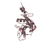
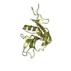
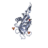
| Title | Crystal structure of calcium2+-free wild-type CD23 lectin domain (crystal form A) | ||||||
 Components Components | Low affinity immunoglobulin epsilon Fc receptor | ||||||
 Keywords Keywords | IMMUNE SYSTEM / immunoglobulin fold lectin / antibody receptor | ||||||
| Function / homology |  Function and homology information Function and homology informationlow-affinity IgE receptor activity / B cell antigen processing and presentation / Fc receptor-mediated immune complex endocytosis / positive regulation of humoral immune response mediated by circulating immunoglobulin / NOTCH2 intracellular domain regulates transcription / macrophage activation / IgE binding / pattern recognition receptor activity / Fc-gamma receptor signaling pathway involved in phagocytosis / Interleukin-10 signaling ...low-affinity IgE receptor activity / B cell antigen processing and presentation / Fc receptor-mediated immune complex endocytosis / positive regulation of humoral immune response mediated by circulating immunoglobulin / NOTCH2 intracellular domain regulates transcription / macrophage activation / IgE binding / pattern recognition receptor activity / Fc-gamma receptor signaling pathway involved in phagocytosis / Interleukin-10 signaling / integrin binding / carbohydrate binding / protease binding / Interleukin-4 and Interleukin-13 signaling / defense response to bacterium / immune response / external side of plasma membrane / positive regulation of gene expression / extracellular exosome / metal ion binding / plasma membrane Similarity search - Function | ||||||
| Biological species |  Homo sapiens (human) Homo sapiens (human) | ||||||
| Method |  X-RAY DIFFRACTION / SYNCHROTRON / MOLECULAR REPLACEMENT / Resolution: 1.9 Å X-RAY DIFFRACTION / SYNCHROTRON / MOLECULAR REPLACEMENT / Resolution: 1.9 Å | ||||||
 Authors Authors | Dhaliwal, B. / Yahya, N. / Sutton, B.J. | ||||||
 Citation Citation | Journal: Mol.Immunol. / Year: 2013 Title: Conformational plasticity at the IgE-binding site of the B-cell receptor CD23. Authors: Dhaliwal, B. / Pang, M.O. / Yuan, D. / Yahya, N. / Fabiane, S.M. / McDonnell, J.M. / Gould, H.J. / Beavil, A.J. / Sutton, B.J. | ||||||
| History |
|
- Structure visualization
| Structure viewer | Molecule: MolmilJmol/JSmol |
|---|
- Downloads & links
-Download
| PDBx/mmCIF format | 4j6j.cif.gz | 232.3 KB | Display | PDBx/mmCIF format |
|---|---|---|---|---|
| PDB format | pdb4j6j.ent.gz | 188 KB | Display | PDB format |
| PDBx/mmJSON format | 4j6j.json.gz | Tree view | PDBx/mmJSON format | |
| Others |  Other downloads Other downloads |
-Validation report
| Arichive directory | https://data.pdbj.org/pub/pdb/validation_reports/j6/4j6jftp://data.pdbj.org/pub/pdb/validation_reports/j6/4j6j | HTTPS FTP |
|---|
-Related structure data
| Related structure data |  4j6kC  4j6lC 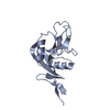 4j6mC  4j6nC  4j6pC  4j6qC  4g96S S: Starting model for refinement C: citing same article ( |
|---|---|
| Similar structure data |


-Links
 PDBj
PDBj


- Assembly
| Deposited unit | 
| ||||||||
|---|---|---|---|---|---|---|---|---|---|
| 1 | 
| ||||||||
| 2 | 
| ||||||||
| 3 | 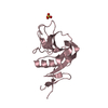
| ||||||||
| 4 | 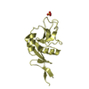
| ||||||||
| Unit cell |
|
-Components
| #1: Protein | Mass: 16164.986 Da / Num. of mol.: 4 Fragment: Soluble head domain of the B-cell receptor CD23 (UNP Residues 156-298) Source method: isolated from a genetically manipulated source Source: (gene. exp.) Homo sapiens (human) / Gene: CD23A, CLEC4J, FCE2, FCER2, IGEBF / Production host:  #2: Chemical |   Mass: 96.063 Da / Num. of mol.: 2 / Source method: obtained synthetically / Formula: SO4 Mass: 96.063 Da / Num. of mol.: 2 / Source method: obtained synthetically / Formula: SO4#3: Water | ChemComp-HOH / |  Mass: 18.015 Da / Num. of mol.: 201 / Source method: isolated from a natural source / Formula: H2O Mass: 18.015 Da / Num. of mol.: 201 / Source method: isolated from a natural source / Formula: H2OHas protein modification | Y | |
|---|
-Experimental details
-Experiment
| Experiment | Method: X-RAY DIFFRACTION / Number of used crystals: 1 |
|---|
- Sample preparation
| Crystal | Density Matthews: 2.58 Å3/Da / Density % sol: 52.25 % |
|---|---|
| Crystal grow | Temperature: 298 K / Method: vapor diffusion, sitting drop / pH: 4.7 Details: 16 % (w/v) PEG 6,000, 2 % (v/v) 1,6-hexanediol, 0.05 M ammonium sulfate and 0.1 M sodium acetate pH 4.7 , VAPOR DIFFUSION, SITTING DROP, temperature 298K |
-Data collection
| Diffraction | Mean temperature: 100 K |
|---|---|
| Diffraction source | Source: SYNCHROTRON / Site: Diamond  / Beamline: I03 / Wavelength: 0.9763 Å / Beamline: I03 / Wavelength: 0.9763 Å |
| Detector | Type: ADSC QUANTUM 315r / Detector: CCD / Date: Jul 10, 2010 |
| Radiation | Protocol: SINGLE WAVELENGTH / Monochromatic (M) / Laue (L): M / Scattering type: x-ray |
| Radiation wavelength | Wavelength: 0.9763 Å / Relative weight: 1 |
| Reflection | Resolution: 1.9→57.96 Å / Num. all: 50712 / Num. obs: 48544 / % possible obs: 96.1 % / Observed criterion σ(F): 0 / Observed criterion σ(I): 0 / Limit h max: 27 / Limit h min: -27 / Limit k max: 29 / Limit k min: -27 / Limit l max: 32 / Limit l min: 0 |
| Reflection shell | % possible obs: 96.3 % / Redundancy: 3.9 % / Rmerge(I) obs: 0.395 / Mean I/σ(I) obs: 2.73 / % possible all: 96.3 |
- Processing
| Software |
| |||||||||||||||||||||||||||||||||||||||||||||||||||||||||||||||||||||||||||||||||||||||||||||||||||||||||||||||||||||||||||||
|---|---|---|---|---|---|---|---|---|---|---|---|---|---|---|---|---|---|---|---|---|---|---|---|---|---|---|---|---|---|---|---|---|---|---|---|---|---|---|---|---|---|---|---|---|---|---|---|---|---|---|---|---|---|---|---|---|---|---|---|---|---|---|---|---|---|---|---|---|---|---|---|---|---|---|---|---|---|---|---|---|---|---|---|---|---|---|---|---|---|---|---|---|---|---|---|---|---|---|---|---|---|---|---|---|---|---|---|---|---|---|---|---|---|---|---|---|---|---|---|---|---|---|---|---|---|---|
| Refinement | Method to determine structure: MOLECULAR REPLACEMENT Starting model: PDB ENTRY 4G96 Resolution: 1.9→15.57 Å / Cor.coef. Fo:Fc: 0.9485 / Cor.coef. Fo:Fc free: 0.9344 / Occupancy max: 1 / Occupancy min: 0.25 / SU R Cruickshank DPI: 0.141 / Cross valid method: THROUGHOUT / σ(F): 0 / Stereochemistry target values: Engh & Huber
| |||||||||||||||||||||||||||||||||||||||||||||||||||||||||||||||||||||||||||||||||||||||||||||||||||||||||||||||||||||||||||||
| Displacement parameters | Biso max: 224.75 Å2 / Biso mean: 40.9941 Å2 / Biso min: 13.41 Å2
| |||||||||||||||||||||||||||||||||||||||||||||||||||||||||||||||||||||||||||||||||||||||||||||||||||||||||||||||||||||||||||||
| Refine analyze | Luzzati coordinate error obs: 0.268 Å | |||||||||||||||||||||||||||||||||||||||||||||||||||||||||||||||||||||||||||||||||||||||||||||||||||||||||||||||||||||||||||||
| Refinement step | Cycle: LAST / Resolution: 1.9→15.57 Å
| |||||||||||||||||||||||||||||||||||||||||||||||||||||||||||||||||||||||||||||||||||||||||||||||||||||||||||||||||||||||||||||
| Refine LS restraints |
| |||||||||||||||||||||||||||||||||||||||||||||||||||||||||||||||||||||||||||||||||||||||||||||||||||||||||||||||||||||||||||||
| LS refinement shell | Resolution: 1.9→1.95 Å / Total num. of bins used: 20
| |||||||||||||||||||||||||||||||||||||||||||||||||||||||||||||||||||||||||||||||||||||||||||||||||||||||||||||||||||||||||||||
| Refinement TLS params. | Method: refined / Refine-ID: X-RAY DIFFRACTION
| |||||||||||||||||||||||||||||||||||||||||||||||||||||||||||||||||||||||||||||||||||||||||||||||||||||||||||||||||||||||||||||
| Refinement TLS group |
|
