 Movie
Movie Controller

[English] 日本語
 Yorodumi
- PDB-4j5n: Crystal Structure of a Deinococcus radiodurans PTE-like lactonase... -
+ Open data

- Basic information
| Entry | Database: PDB / ID: 4j5n | ||||||
|---|---|---|---|---|---|---|---|
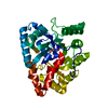
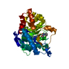
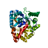
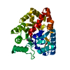
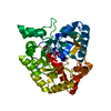
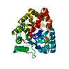
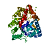
| Title | Crystal Structure of a Deinococcus radiodurans PTE-like lactonase (drPLL) mutant Y28L/D71N/E101G/E179D/V235L/L270M | ||||||
 Components Components | Phosphotriesterase, putative | ||||||
 Keywords Keywords | HYDROLASE / Metalloenzyme / Tim Barrel / Nerve agent / Phosphotriesterase / Lactonase | ||||||
| Function / homology |  Function and homology information Function and homology information | ||||||
| Biological species |  Deinococcus radiodurans (radioresistant) Deinococcus radiodurans (radioresistant) | ||||||
| Method |  X-RAY DIFFRACTION / SYNCHROTRON / MOLECULAR REPLACEMENT / Resolution: 2.05 Å X-RAY DIFFRACTION / SYNCHROTRON / MOLECULAR REPLACEMENT / Resolution: 2.05 Å | ||||||
 Authors Authors | Meier, M.M. / Rajendran, C. / Malisi, C. / Fox, N.G. / Xu, C. / Schlee, S. / Barondeau, D.P. / Hocker, B. / Sterner, R. / Raushel, F.M. | ||||||
 Citation Citation | Journal: J.Am.Chem.Soc. / Year: 2013 Title: Molecular engineering of organophosphate hydrolysis activity from a weak promiscuous lactonase template. Authors: Meier, M.M. / Rajendran, C. / Malisi, C. / Fox, N.G. / Xu, C. / Schlee, S. / Barondeau, D.P. / Hocker, B. / Sterner, R. / Raushel, F.M. | ||||||
| History |
|
- Structure visualization
| Structure viewer | Molecule: MolmilJmol/JSmol |
|---|
- Downloads & links
-Download
| PDBx/mmCIF format | 4j5n.cif.gz | 80.2 KB | Display | PDBx/mmCIF format |
|---|---|---|---|---|
| PDB format | pdb4j5n.ent.gz | 58 KB | Display | PDB format |
| PDBx/mmJSON format | 4j5n.json.gz | Tree view | PDBx/mmJSON format | |
| Others |  Other downloads Other downloads |
-Validation report
| Arichive directory | https://data.pdbj.org/pub/pdb/validation_reports/j5/4j5nftp://data.pdbj.org/pub/pdb/validation_reports/j5/4j5n | HTTPS FTP |
|---|
-Related structure data
| Related structure data |  4j2mC  4j35C  3fdkS S: Starting model for refinement C: citing same article ( |
|---|---|
| Similar structure data |


-Links
 PDBj
PDBj


- Assembly
| Deposited unit | 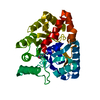
| ||||||||
|---|---|---|---|---|---|---|---|---|---|
| 1 |
| ||||||||
| 2 | 
| ||||||||
| Unit cell |
|
-Components
| #1: Protein | Mass: 34704.102 Da / Num. of mol.: 1 / Mutation: Y28L, D71N, E101G, E179D, V235L, L270M Source method: isolated from a genetically manipulated source Source: (gene. exp.) Deinococcus radiodurans (radioresistant)Gene: Dr0930 (gi 15805954), DR_0930 / Production host: | ||
|---|---|---|---|
| #2: Chemical |   Mass: 58.933 Da / Num. of mol.: 2 / Source method: obtained synthetically / Formula: Co Mass: 58.933 Da / Num. of mol.: 2 / Source method: obtained synthetically / Formula: Co#3: Water | ChemComp-HOH / |  Mass: 18.015 Da / Num. of mol.: 210 / Source method: isolated from a natural source / Formula: H2O Mass: 18.015 Da / Num. of mol.: 210 / Source method: isolated from a natural source / Formula: H2O |
-Experimental details
-Experiment
| Experiment | Method: X-RAY DIFFRACTION / Number of used crystals: 1 |
|---|
- Sample preparation
| Crystal | Density Matthews: 3.22 Å3/Da / Density % sol: 61.8 % |
|---|---|
| Crystal grow | Temperature: 295 K / Method: vapor diffusion, hanging drop / pH: 8 Details: 2 uL of protein (~10 mg/mL) and 2 uL of well solution (0.1 M imidazole pH 8.0, 10% PEG 8000, 0.2 M Ca(OAc)2 ) were mixed and placed on a siliconized coverslip over 500 uL of the well ...Details: 2 uL of protein (~10 mg/mL) and 2 uL of well solution (0.1 M imidazole pH 8.0, 10% PEG 8000, 0.2 M Ca(OAc)2 ) were mixed and placed on a siliconized coverslip over 500 uL of the well solution. Cryo 14% EG, VAPOR DIFFUSION, HANGING DROP, temperature 295K |
-Data collection
| Diffraction | Mean temperature: 100 K |
|---|---|
| Diffraction source | Source: SYNCHROTRON / Site: SSRL  / Beamline: BL7-1 / Wavelength: 0.9794 Å / Beamline: BL7-1 / Wavelength: 0.9794 Å |
| Detector | Type: ADSC QUANTUM 315r / Detector: CCD / Date: Jul 23, 2010 |
| Radiation | Monochromator: Si(111), side scattering I-beam bent single crystal; asymmetric cut 4.9650 deg Protocol: SINGLE WAVELENGTH / Monochromatic (M) / Laue (L): M / Scattering type: x-ray |
| Radiation wavelength | Wavelength: 0.9794 Å / Relative weight: 1 |
| Reflection | Resolution: 2.05→50 Å / Num. obs: 29424 / % possible obs: 100 % / Observed criterion σ(F): 0 / Observed criterion σ(I): 0 / Redundancy: 18.4 % / Biso Wilson estimate: 38.6 Å2 / Rsym value: 0.137 / Net I/σ(I): 36.5 |
| Reflection shell | Resolution: 2.05→2.09 Å / Redundancy: 19.1 % / Mean I/σ(I) obs: 7.8 / Num. unique all: 1417 / Rsym value: 0.473 / % possible all: 100 |
- Processing
| Software |
| |||||||||||||||||||||||||
|---|---|---|---|---|---|---|---|---|---|---|---|---|---|---|---|---|---|---|---|---|---|---|---|---|---|---|
| Refinement | Method to determine structure: MOLECULAR REPLACEMENT Starting model: 3FDK Resolution: 2.05→50 Å / Cross valid method: THROUGHOUT / σ(F): 0 / σ(I): 0 / Stereochemistry target values: Engh & Huber
| |||||||||||||||||||||||||
| Refinement step | Cycle: LAST / Resolution: 2.05→50 Å
| |||||||||||||||||||||||||
| Refine LS restraints |
| |||||||||||||||||||||||||
| LS refinement shell | Resolution: 2.05→2.07 Å /
|
