 Movie
Movie Controller

[English] 日本語
 Yorodumi
- PDB-4j3o: Crystal structure of the FimD usher traversed by the pilus tip co... -
+ Open data

- Basic information
| Entry | Database: PDB / ID: 4j3o | ||||||
|---|---|---|---|---|---|---|---|
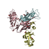
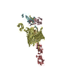
| Title | Crystal structure of the FimD usher traversed by the pilus tip complex assembly composed of FimC:FimF:FimG:FimH | ||||||
 Components Components |
| ||||||
 Keywords Keywords | CELL ADHESION/CHAPERONE/MEMBRANE PROTEIN / beta barrel / immunglobuline-like fold / type 1 pilus assembly / pilus subunit translocation / adhesion / D-Mannose-binding / bacterial outer membrane / CELL ADHESION-CHAPERONE-MEMBRANE PROTEIN complex | ||||||
| Function / homology |  Function and homology information Function and homology informationfimbrial usher porin activity / pilus assembly / pilus tip / mechanosensory behavior / Attachment of bacteria to epithelial cells / cell adhesion involved in single-species biofilm formation / pilus / cell-substrate adhesion / D-mannose binding / host cell membrane ...fimbrial usher porin activity / pilus assembly / pilus tip / mechanosensory behavior / Attachment of bacteria to epithelial cells / cell adhesion involved in single-species biofilm formation / pilus / cell-substrate adhesion / D-mannose binding / host cell membrane / protein folding chaperone / cell outer membrane / cell wall organization / outer membrane-bounded periplasmic space / protein folding / cell adhesion Similarity search - Function | ||||||
| Biological species |  | ||||||
| Method |  X-RAY DIFFRACTION / SYNCHROTRON / MOLECULAR REPLACEMENT / Resolution: 3.8 Å X-RAY DIFFRACTION / SYNCHROTRON / MOLECULAR REPLACEMENT / Resolution: 3.8 Å | ||||||
 Authors Authors | Geibel, S. / Waksman, G. | ||||||
 Citation Citation | Journal: Nature / Year: 2013 Title: Structural and energetic basis of folded-protein transport by the FimD usher. Authors: Geibel, S. / Procko, E. / Hultgren, S.J. / Baker, D. / Waksman, G. | ||||||
| History |
|
- Structure visualization
| Structure viewer | Molecule: MolmilJmol/JSmol |
|---|
- Downloads & links
-Download
| PDBx/mmCIF format | 4j3o.cif.gz | 308.1 KB | Display | PDBx/mmCIF format |
|---|---|---|---|---|
| PDB format | pdb4j3o.ent.gz | 246.1 KB | Display | PDB format |
| PDBx/mmJSON format | 4j3o.json.gz | Tree view | PDBx/mmJSON format | |
| Others |  Other downloads Other downloads |
-Validation report
| Arichive directory | https://data.pdbj.org/pub/pdb/validation_reports/j3/4j3oftp://data.pdbj.org/pub/pdb/validation_reports/j3/4j3o | HTTPS FTP |
|---|
-Related structure data


-Links
 PDBj
PDBj- Assembly
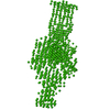
| Deposited unit | 
| ||||||||
|---|---|---|---|---|---|---|---|---|---|
| 1 |
| ||||||||
| Unit cell |
|
-Components
| #1: Protein | Mass: 14864.227 Da / Num. of mol.: 1 / Fragment: UNP residues 24-167 Source method: isolated from a genetically manipulated source Source: (gene. exp.) |
|---|---|
| #2: Protein | Mass: 29081.314 Da / Num. of mol.: 1 / Fragment: UNP residues 22-300 Source method: isolated from a genetically manipulated source Source: (gene. exp.) |
| #3: Protein | Mass: 23582.914 Da / Num. of mol.: 1 / Fragment: UNP residues 37-241 Source method: isolated from a genetically manipulated source Source: (gene. exp.) |
| #4: Protein | Mass: 16177.095 Da / Num. of mol.: 1 / Fragment: UNP residues 21-176 Source method: isolated from a genetically manipulated source Source: (gene. exp.) |
| #5: Protein | Mass: 92640.227 Da / Num. of mol.: 1 / Fragment: UNP residues 46-878 Source method: isolated from a genetically manipulated source Source: (gene. exp.) |
| Has protein modification | Y |
-Experimental details
-Experiment
| Experiment | Method: X-RAY DIFFRACTION / Number of used crystals: 1 |
|---|
- Sample preparation
| Crystal | Density Matthews: 3.48 Å3/Da / Density % sol: 64.68 % |
|---|---|
| Crystal grow | Temperature: 298 K / Method: vapor diffusion, hanging drop / pH: 8.5 Details: 1.6-2.0M sodium formate, pH 8.5, VAPOR DIFFUSION, HANGING DROP, temperature 298K |
-Data collection
| Diffraction | Mean temperature: 100 K |
|---|---|
| Diffraction source | Source: SYNCHROTRON / Site: Diamond  / Beamline: I04 / Wavelength: 0.9795 Å / Beamline: I04 / Wavelength: 0.9795 Å |
| Detector | Type: ADSC QUANTUM 315r / Detector: CCD / Date: Dec 6, 2010 / Details: mirrors |
| Radiation | Monochromator: Si111 double crystal monochromator / Protocol: SINGLE WAVELENGTH / Monochromatic (M) / Laue (L): M / Scattering type: x-ray |
| Radiation wavelength | Wavelength: 0.9795 Å / Relative weight: 1 |
| Reflection | Resolution: 3.8→83.67 Å / Num. all: 25518 / Num. obs: 25474 / % possible obs: 99.84 % / Observed criterion σ(I): -3 |
| Reflection shell | Resolution: 3.8→3.9 Å / % possible all: 99.96 |
- Processing
| Software |
| ||||||||||||||||||||||||||||||||||||||||||||||||||||||||||||||||||||||
|---|---|---|---|---|---|---|---|---|---|---|---|---|---|---|---|---|---|---|---|---|---|---|---|---|---|---|---|---|---|---|---|---|---|---|---|---|---|---|---|---|---|---|---|---|---|---|---|---|---|---|---|---|---|---|---|---|---|---|---|---|---|---|---|---|---|---|---|---|---|---|---|
| Refinement | Method to determine structure: MOLECULAR REPLACEMENT Starting model: PDB entry 3BWU for FimC:FimF, PDB 3RFZ for FimD, PDB 3JWN for FimG:FimH Resolution: 3.8→83.668 Å / SU ML: 0.76 / σ(F): 1.33 / Phase error: 37.86 / Stereochemistry target values: ML
| ||||||||||||||||||||||||||||||||||||||||||||||||||||||||||||||||||||||
| Solvent computation | Shrinkage radii: 0.9 Å / VDW probe radii: 1.11 Å / Solvent model: FLAT BULK SOLVENT MODEL | ||||||||||||||||||||||||||||||||||||||||||||||||||||||||||||||||||||||
| Refinement step | Cycle: LAST / Resolution: 3.8→83.668 Å
| ||||||||||||||||||||||||||||||||||||||||||||||||||||||||||||||||||||||
| Refine LS restraints |
| ||||||||||||||||||||||||||||||||||||||||||||||||||||||||||||||||||||||
| LS refinement shell |
|
