 Movie
Movie Controller


+ Open data

- Basic information
| Entry | Database: PDB / ID: 4j2n | ||||||
|---|---|---|---|---|---|---|---|
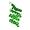
| Title | Crystal Structure of mycobacteriophage Pukovnik Xis | ||||||
 Components Components | Gp37 | ||||||
 Keywords Keywords | VIRAL PROTEIN / Winged-Helix / Doman Swap / Filament | ||||||
| Function / homology | SinI-like, DNA-binding domain / Helix-turn-helix domain, group 17 / Helix-turn-helix domain / Putative DNA-binding domain superfamily / DNA binding / Excise Function and homology information Function and homology information | ||||||
| Biological species |  Mycobacterium phage Pukovnik (virus) Mycobacterium phage Pukovnik (virus) | ||||||
| Method |  X-RAY DIFFRACTION / SYNCHROTRON / SAD / Resolution: 2.348 Å X-RAY DIFFRACTION / SYNCHROTRON / SAD / Resolution: 2.348 Å | ||||||
 Authors Authors | Homa, N.J. / Amrich, C.G. / Heroux, A. / VanDemark, A.P. | ||||||
 Citation Citation | Journal: J.Mol.Biol. / Year: 2014 Title: The Structure of Xis Reveals the Basis for Filament Formation and Insight into DNA Bending within a Mycobacteriophage Intasome. Authors: Singh, S. / Plaks, J.G. / Homa, N.J. / Amrich, C.G. / Heroux, A. / Hatfull, G.F. / Vandemark, A.P. | ||||||
| History |
|
- Structure visualization
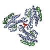
| Structure viewer | Molecule: MolmilJmol/JSmol |
|---|
- Downloads & links
-Download
| PDBx/mmCIF format | 4j2n.cif.gz | 67 KB | Display | PDBx/mmCIF format |
|---|---|---|---|---|
| PDB format | pdb4j2n.ent.gz | 51.3 KB | Display | PDB format |
| PDBx/mmJSON format | 4j2n.json.gz | Tree view | PDBx/mmJSON format | |
| Others |  Other downloads Other downloads |
-Validation report
| Arichive directory | https://data.pdbj.org/pub/pdb/validation_reports/j2/4j2nftp://data.pdbj.org/pub/pdb/validation_reports/j2/4j2n | HTTPS FTP |
|---|
-Related structure data
| Similar structure data |
|---|


-Links
 PDBj
PDBj

- Assembly
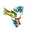
| Deposited unit | 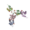
| ||||||||
|---|---|---|---|---|---|---|---|---|---|
| 1 |
| ||||||||
| Unit cell |
|
-Components
| #1: Protein | Mass: 6406.617 Da / Num. of mol.: 5 Source method: isolated from a genetically manipulated source Source: (gene. exp.) Mycobacterium phage Pukovnik (virus) / Strain: Pukovnik / Gene: 37, Pukovnik_37, XIS / Plasmid: pMCSG7 / Production host:  #2: Chemical | ChemComp-SO4 /   Mass: 96.063 Da / Num. of mol.: 8 / Source method: obtained synthetically / Formula: SO4 Mass: 96.063 Da / Num. of mol.: 8 / Source method: obtained synthetically / Formula: SO4#3: Water | ChemComp-HOH / |  Mass: 18.015 Da / Num. of mol.: 140 / Source method: isolated from a natural source / Formula: H2O Mass: 18.015 Da / Num. of mol.: 140 / Source method: isolated from a natural source / Formula: H2O |
|---|
-Experimental details
-Experiment
| Experiment | Method: X-RAY DIFFRACTION / Number of used crystals: 2 |
|---|
- Sample preparation
| Crystal | Density Matthews: 4.19 Å3/Da / Density % sol: 70.63 % |
|---|---|
| Crystal grow | Temperature: 300 K / Method: vapor diffusion, sitting drop / pH: 8 Details: ammonium sulfate, PEG 3350, pH 8.0, VAPOR DIFFUSION, SITTING DROP, temperature 300K |
-Data collection
| Diffraction |
| ||||||||||||||||||
|---|---|---|---|---|---|---|---|---|---|---|---|---|---|---|---|---|---|---|---|
| Diffraction source |
| ||||||||||||||||||
| Detector |
| ||||||||||||||||||
| Radiation |
| ||||||||||||||||||
| Radiation wavelength |
| ||||||||||||||||||
| Reflection | Resolution: 2.35→90 Å / Num. all: 22860 / Num. obs: 22815 / % possible obs: 99.8 % / Observed criterion σ(F): 3 / Observed criterion σ(I): 2 / Biso Wilson estimate: 45.18 Å2 | ||||||||||||||||||
| Reflection shell | Resolution: 2.35→2.39 Å / Redundancy: 4.2 % / Mean I/σ(I) obs: 2.2 / Num. unique all: 1127 / % possible all: 100 |

- Processing
| Software |
| |||||||||||||||||||||||||||||||||||||||||||||||||||||||||||||||||||||||||||||
|---|---|---|---|---|---|---|---|---|---|---|---|---|---|---|---|---|---|---|---|---|---|---|---|---|---|---|---|---|---|---|---|---|---|---|---|---|---|---|---|---|---|---|---|---|---|---|---|---|---|---|---|---|---|---|---|---|---|---|---|---|---|---|---|---|---|---|---|---|---|---|---|---|---|---|---|---|---|---|
| Refinement | Method to determine structure: SAD / Resolution: 2.348→35.921 Å / Occupancy max: 1 / Occupancy min: 0.39 / FOM work R set: 0.7672 / SU ML: 0.35 / σ(F): 0.03 / Phase error: 29.32 / Stereochemistry target values: ML
| |||||||||||||||||||||||||||||||||||||||||||||||||||||||||||||||||||||||||||||
| Solvent computation | Shrinkage radii: 0.9 Å / VDW probe radii: 1.11 Å / Solvent model: FLAT BULK SOLVENT MODEL / Bsol: 60.428 Å2 / ksol: 0.392 e/Å3 | |||||||||||||||||||||||||||||||||||||||||||||||||||||||||||||||||||||||||||||
| Displacement parameters | Biso max: 112.21 Å2 / Biso mean: 54.6141 Å2 / Biso min: 34.42 Å2
| |||||||||||||||||||||||||||||||||||||||||||||||||||||||||||||||||||||||||||||
| Refinement step | Cycle: LAST / Resolution: 2.348→35.921 Å
| |||||||||||||||||||||||||||||||||||||||||||||||||||||||||||||||||||||||||||||
| Refine LS restraints |
| |||||||||||||||||||||||||||||||||||||||||||||||||||||||||||||||||||||||||||||
| LS refinement shell | Refine-ID: X-RAY DIFFRACTION / Total num. of bins used: 10
|
