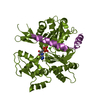
 Movie
Movie Controller


+ Open data

- Basic information
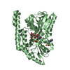
| Entry | Database: PDB / ID: 4iu9 | ||||||
|---|---|---|---|---|---|---|---|
| Title | Crystal structure of a membrane transporter | ||||||
 Components Components | Nitrite extrusion protein 2 | ||||||
 Keywords Keywords | TRANSPORT PROTEIN / membrane protein / nitrate-nitrite porter family transporter / MFS fold | ||||||
| Function / homology |  Function and homology information Function and homology informationnitrite efflux transmembrane transporter activity / nitrite transport / nitrate transmembrane transporter activity / nitrate transmembrane transport / nitrate assimilation / plasma membrane Similarity search - Function | ||||||
| Biological species |  | ||||||
| Method |  X-RAY DIFFRACTION / SYNCHROTRON / MOLECULAR REPLACEMENT / Resolution: 3.005 Å X-RAY DIFFRACTION / SYNCHROTRON / MOLECULAR REPLACEMENT / Resolution: 3.005 Å | ||||||
 Authors Authors | Yan, H. / Huang, W. / Yan, C. / Gong, X. / Jiang, S. / Zhao, Y. / Wang, J. / Shi, Y. | ||||||
 Citation Citation | Journal: Cell Rep / Year: 2013 Title: Structure and mechanism of a nitrate transporter. Authors: Yan, H. / Huang, W. / Yan, C. / Gong, X. / Jiang, S. / Zhao, Y. / Wang, J. / Shi, Y. | ||||||
| History |
|
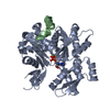
- Structure visualization
| Structure viewer | Molecule: MolmilJmol/JSmol |
|---|
- Downloads & links
-Download
| PDBx/mmCIF format | 4iu9.cif.gz | 306.9 KB | Display | PDBx/mmCIF format |
|---|---|---|---|---|
| PDB format | pdb4iu9.ent.gz | 255.8 KB | Display | PDB format |
| PDBx/mmJSON format | 4iu9.json.gz | Tree view | PDBx/mmJSON format | |
| Others |  Other downloads Other downloads |
-Validation report
| Summary document | 4iu9_validation.pdf.gz | 441.8 KB | Display | wwPDB validaton report |
|---|---|---|---|---|
| Full document | 4iu9_full_validation.pdf.gz | 461 KB | Display | |
| Data in XML | 4iu9_validation.xml.gz | 29.6 KB | Display | |
| Data in CIF | 4iu9_validation.cif.gz | 40.4 KB | Display | |
| Arichive directory | https://data.pdbj.org/pub/pdb/validation_reports/iu/4iu9ftp://data.pdbj.org/pub/pdb/validation_reports/iu/4iu9 | HTTPS FTP |
-Related structure data
| Related structure data | 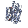 4iu8SC S: Starting model for refinement C: citing same article ( |
|---|---|
| Similar structure data |


-Links
 PDBj
PDBj


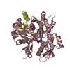
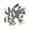
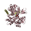
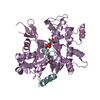
- Assembly
| Deposited unit | 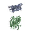
| ||||||||
|---|---|---|---|---|---|---|---|---|---|
| 1 | 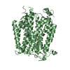
| ||||||||
| 2 | 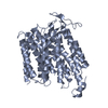
| ||||||||
| Unit cell |
|
-Components
| #1: Protein | Mass: 50653.637 Da / Num. of mol.: 2 Source method: isolated from a genetically manipulated source Source: (gene. exp.) |
|---|
-Experimental details
-Experiment
| Experiment | Method: X-RAY DIFFRACTION / Number of used crystals: 1 |
|---|
- Sample preparation
| Crystal | Density Matthews: 4.42 Å3/Da / Density % sol: 72.15 % |
|---|---|
| Crystal grow | Temperature: 291 K / Method: vapor diffusion, hanging drop / pH: 8.5 Details: 100 mM Tris-HCl (pH 8.5), 33% (w/v) PEG 400 and 100 mM KCl, VAPOR DIFFUSION, HANGING DROP, temperature 291K |
-Data collection
| Diffraction | Mean temperature: 100 K |
|---|---|
| Diffraction source | Source: SYNCHROTRON / Site: SSRF  / Beamline: BL17U / Wavelength: 1.07082 Å / Beamline: BL17U / Wavelength: 1.07082 Å |
| Detector | Type: ADSC QUANTUM 315r / Detector: CCD / Date: Nov 20, 2011 |
| Radiation | Monochromator: Si 111 CHANNEL / Protocol: SINGLE WAVELENGTH / Monochromatic (M) / Laue (L): M / Scattering type: x-ray |
| Radiation wavelength | Wavelength: 1.07082 Å / Relative weight: 1 |
| Reflection | Resolution: 3→40 Å / Num. all: 35777 / Num. obs: 30017 / % possible obs: 83.9 % / Observed criterion σ(I): -3 |
| Reflection shell | Resolution: 3→3.11 Å / % possible all: 62.2 |
- Processing
| Software |
| |||||||||||||||||||||||||||||||||||||||||||||||||||||||||||||||||||||||||||||
|---|---|---|---|---|---|---|---|---|---|---|---|---|---|---|---|---|---|---|---|---|---|---|---|---|---|---|---|---|---|---|---|---|---|---|---|---|---|---|---|---|---|---|---|---|---|---|---|---|---|---|---|---|---|---|---|---|---|---|---|---|---|---|---|---|---|---|---|---|---|---|---|---|---|---|---|---|---|---|
| Refinement | Method to determine structure: MOLECULAR REPLACEMENT Starting model: 4IU8 Resolution: 3.005→39.84 Å / SU ML: 0.35 / Cross valid method: THROUGHOUT / σ(F): 1.33 / Phase error: 39.81 / Stereochemistry target values: ML
| |||||||||||||||||||||||||||||||||||||||||||||||||||||||||||||||||||||||||||||
| Solvent computation | Shrinkage radii: 0.47 Å / VDW probe radii: 0.8 Å / Solvent model: FLAT BULK SOLVENT MODEL / Bsol: 84.45 Å2 / ksol: 0.286 e/Å3 | |||||||||||||||||||||||||||||||||||||||||||||||||||||||||||||||||||||||||||||
| Displacement parameters |
| |||||||||||||||||||||||||||||||||||||||||||||||||||||||||||||||||||||||||||||
| Refinement step | Cycle: LAST / Resolution: 3.005→39.84 Å
| |||||||||||||||||||||||||||||||||||||||||||||||||||||||||||||||||||||||||||||
| Refine LS restraints |
| |||||||||||||||||||||||||||||||||||||||||||||||||||||||||||||||||||||||||||||
| LS refinement shell |
| |||||||||||||||||||||||||||||||||||||||||||||||||||||||||||||||||||||||||||||
| Refinement TLS params. | Method: refined / Origin x: -0.4538 Å / Origin y: -1.0669 Å / Origin z: -17.9241 Å
| |||||||||||||||||||||||||||||||||||||||||||||||||||||||||||||||||||||||||||||
| Refinement TLS group | Selection details: all |
