- PDB-4ip9: Structure of native human serum amyloid A1 -
+
Open data
ID or keywords:
Loading...
-
Basic information
Entry
Database: PDB / ID: 4ip9
Title
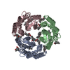
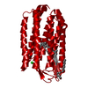
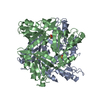
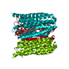
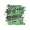
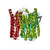
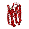
Structure of native human serum amyloid A1
Components
Serum amyloid A-1 protein
Keywords
PROTEIN BINDING / double layer hexameric structure / secondary amyloid / High density lipoprotein / human serum
Function / homology
Function and homology information
lymphocyte chemotaxis / Scavenging by Class B Receptors / positive regulation of interleukin-1 production / high-density lipoprotein particle / Formyl peptide receptors bind formyl peptides and many other ligands / regulation of protein secretion / TRAF6 mediated NF-kB activation / macrophage chemotaxis / Advanced glycosylation endproduct receptor signaling / cytoplasmic microtubule ...lymphocyte chemotaxis / Scavenging by Class B Receptors / positive regulation of interleukin-1 production / high-density lipoprotein particle / Formyl peptide receptors bind formyl peptides and many other ligands / regulation of protein secretion / TRAF6 mediated NF-kB activation / macrophage chemotaxis / Advanced glycosylation endproduct receptor signaling / cytoplasmic microtubule / neutrophil chemotaxis / endocytic vesicle lumen / positive regulation of cell adhesion / positive regulation of cytokine production / acute-phase response / platelet activation / TAK1-dependent IKK and NF-kappa-B activation / G protein-coupled receptor binding / negative regulation of inflammatory response / heparin binding / positive regulation of cytosolic calcium ion concentration / Interleukin-4 and Interleukin-13 signaling / G alpha (i) signalling events / G alpha (q) signalling events / Amyloid fiber formation / extracellular exosome / extracellular region Similarity search - Function
Serum amyloid A protein / Serum amyloid A protein / : / Serum amyloid A protein / Serum amyloid A proteins signature. / Serum amyloid A proteins / Topoisomerase I; Chain A, domain 4 / Orthogonal Bundle / Mainly Alpha Similarity search - Domain/homology
SerumamyloidA-1protein / SAA / Amyloid protein A / Amyloid fibril protein AA / Serum amyloid protein A(2-104) / Serum ...SAA / Amyloid protein A / Amyloid fibril protein AA / Serum amyloid protein A(2-104) / Serum amyloid protein A(3-104) / Serum amyloid protein A(2-103) / Serum amyloid protein A(2-102) / Serum amyloid protein A(4-101)
Mass: 12659.771 Da / Num. of mol.: 2 Source method: isolated from a genetically manipulated source Source: (gene. exp.) Homo sapiens (human) / Gene: SAA1 / Production host: Escherichia coli (E. coli) / References: UniProt: P0DJI8
Mass: 18.015 Da / Num. of mol.: 6 / Source method: isolated from a natural source / Formula: H2O
-
Experimental details
-
Experiment
Experiment
Method: X-RAY DIFFRACTION
-
Sample preparation
Crystal
Density Matthews: 2.18 Å3/Da / Density % sol: 43.69 %
Crystal grow
Temperature: 295 K / Method: vapor diffusion, hanging drop / pH: 8.5 Details: 30% PEG2000 MME, 0.1M Tris (pH8.5) and 0.2 M Trimethylamine N-oxide, VAPOR DIFFUSION, HANGING DROP, temperature 295K
-
Data collection
Diffraction
Mean temperature: 100 K
Diffraction source
Source: SYNCHROTRON / Site: APS / Beamline: 22-ID
Detector
Type: MAR CCD 165 mm / Detector: CCD / Date: Jul 4, 2010
Radiation
Protocol: SINGLE WAVELENGTH / Monochromatic (M) / Laue (L): M / Scattering type: x-ray
In the structure databanks used in Yorodumi, some data are registered as the other names, "COVID-19 virus" and "2019-nCoV". Here are the details of the virus and the list of structure data.
Jan 31, 2019. EMDB accession codes are about to change! (news from PDBe EMDB page)
EMDB accession codes are about to change! (news from PDBe EMDB page)
The allocation of 4 digits for EMDB accession codes will soon come to an end. Whilst these codes will remain in use, new EMDB accession codes will include an additional digit and will expand incrementally as the available range of codes is exhausted. The current 4-digit format prefixed with “EMD-” (i.e. EMD-XXXX) will advance to a 5-digit format (i.e. EMD-XXXXX), and so on. It is currently estimated that the 4-digit codes will be depleted around Spring 2019, at which point the 5-digit format will come into force.
The EM Navigator/Yorodumi systems omit the EMD- prefix.
Related info.:Q: What is EMD? / ID/Accession-code notation in Yorodumi/EM Navigator
Yorodumi is a browser for structure data from EMDB, PDB, SASBDB, etc.
This page is also the successor to EM Navigator detail page, and also detail information page/front-end page for Omokage search.
The word "yorodu" (or yorozu) is an old Japanese word meaning "ten thousand". "mi" (miru) is to see.
Related info.:EMDB / PDB / SASBDB / Comparison of 3 databanks / Yorodumi Search / Aug 31, 2016. New EM Navigator & Yorodumi / Yorodumi Papers / Jmol/JSmol / Function and homology information / Changes in new EM Navigator and Yorodumi
 Movie
Movie Controller


 Open data
 Basic information
 Components
 Keywords
 Function and homology information
 Homo sapiens (human)
 X-RAY DIFFRACTION /
  Authors
 Citation
 Structure visualization
 Downloads & links
 Other downloads

 PDBj
PDBj


 Assembly
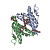


 Mass: 324.367 Da / Num. of mol.: 6 / Source method: obtained synthetically / Formula: C14H28O8
 Mass: 18.015 Da / Num. of mol.: 6 / Source method: isolated from a natural source / Formula: H2O
 Sample preparation
 / Beamline: 22-ID
 Processing