 Movie
Movie Controller

[English] 日本語
 Yorodumi
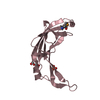
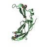
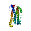
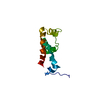
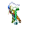
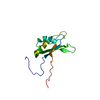
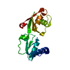
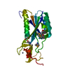
- PDB-4im9: Cystal structure of DnaG primase C-terminal domain from Vibrio ch... -
+ Open data

- Basic information
| Entry | Database: PDB / ID: 4im9 | ||||||
|---|---|---|---|---|---|---|---|
| Title | Cystal structure of DnaG primase C-terminal domain from Vibrio cholerae | ||||||
 Components Components | DNA primase | ||||||
 Keywords Keywords | TRANSFERASE / helicase-primase complex / DNA replication / Hair pin helix / helicase binding | ||||||
| Function / homology | DNAb Helicase; Chain A / DNAb Helicase; Chain A / Orthogonal Bundle / Mainly Alpha / :  Function and homology information Function and homology information | ||||||
| Biological species |   Vibrio cholerae (bacteria) Vibrio cholerae (bacteria) | ||||||
| Method |  X-RAY DIFFRACTION / SYNCHROTRON / SAD / Resolution: 2.46 Å X-RAY DIFFRACTION / SYNCHROTRON / SAD / Resolution: 2.46 Å | ||||||
 Authors Authors | Abdul Rehman, S.A. / Tarique, K.F. / Gourinath, S. | ||||||
 Citation Citation | Journal: To be Published Title: Cystal structure of DnaG primase C-terminal domain from Vibrio cholerae Authors: Abdul Rehman, S.A. / Tarique, K.F. / Gourinath, S. | ||||||
| History |
|
- Structure visualization
| Structure viewer | Molecule: MolmilJmol/JSmol |
|---|
- Downloads & links
-Download
| PDBx/mmCIF format | 4im9.cif.gz | 91.9 KB | Display | PDBx/mmCIF format |
|---|---|---|---|---|
| PDB format | pdb4im9.ent.gz | 71.3 KB | Display | PDB format |
| PDBx/mmJSON format | 4im9.json.gz | Tree view | PDBx/mmJSON format | |
| Others |  Other downloads Other downloads |
-Validation report
| Arichive directory | https://data.pdbj.org/pub/pdb/validation_reports/im/4im9ftp://data.pdbj.org/pub/pdb/validation_reports/im/4im9 | HTTPS FTP |
|---|
-Related structure data
| Similar structure data |
|---|


-Links
 PDBj
PDBj- Assembly
| Deposited unit | 
| ||||||||
|---|---|---|---|---|---|---|---|---|---|
| 1 | 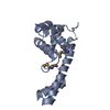
| ||||||||
| 2 | 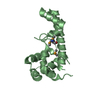
| ||||||||
| 3 | 
| ||||||||
| Unit cell |
|
-Components
| #1: Protein | Mass: 17706.951 Da / Num. of mol.: 3 / Fragment: Primase C-terminal domain Source method: isolated from a genetically manipulated source Source: (gene. exp.) Vibrio cholerae (bacteria) / Strain: O395 / Gene: C-terminal domain, dnaG, Primase, VC395_0535 / Plasmid: pET28(b) / Production host: References: UniProt: C3LX44, Transferases; Transferring phosphorus-containing groups; Nucleotidyltransferases #2: Water | ChemComp-HOH / |  Mass: 18.015 Da / Num. of mol.: 43 / Source method: isolated from a natural source / Formula: H2O Mass: 18.015 Da / Num. of mol.: 43 / Source method: isolated from a natural source / Formula: H2OHas protein modification | Y | |
|---|
-Experimental details
-Experiment
| Experiment | Method: X-RAY DIFFRACTION / Number of used crystals: 1 |
|---|
- Sample preparation
| Crystal | Density Matthews: 3.15 Å3/Da / Density % sol: 60.97 % |
|---|---|
| Crystal grow | Temperature: 289 K / Method: vapor diffusion, hanging drop / pH: 6.5 Details: 0.1 M Sodium cacodylate trihydrate (pH 6.5), 0.2 M MgCl2, 30% W/v PEG 3350 , VAPOR DIFFUSION, HANGING DROP, temperature 289K |
-Data collection
| Diffraction | Mean temperature: 100 K |
|---|---|
| Diffraction source | Source: SYNCHROTRON / Site: ESRF  / Beamline: BM14 / Wavelength: 0.97872 Å / Beamline: BM14 / Wavelength: 0.97872 Å |
| Detector | Type: MAR scanner 345 mm plate / Detector: IMAGE PLATE / Date: May 21, 2012 / Details: bent collimating mirror and toroid |
| Radiation | Monochromator: Si(111) monochromator / Protocol: SINGLE WAVELENGTH / Monochromatic (M) / Laue (L): M / Scattering type: x-ray |
| Radiation wavelength | Wavelength: 0.97872 Å / Relative weight: 1 |
| Reflection | Resolution: 2.45→66.326 Å / Num. all: 25083 / Num. obs: 25046 / % possible obs: 99.9 % / Observed criterion σ(F): 0 / Observed criterion σ(I): -3 / Redundancy: 4.4 % / Rmerge(I) obs: 0.074 |
| Reflection shell | Resolution: 2.45→2.49 Å / Redundancy: 4.1 % / Rmerge(I) obs: 0.404 / Mean I/σ(I) obs: 2.7 / Num. unique all: 1189 / % possible all: 98.3 |
- Processing
| Software |
| |||||||||||||||||||||||||||||||||||||||||||||
|---|---|---|---|---|---|---|---|---|---|---|---|---|---|---|---|---|---|---|---|---|---|---|---|---|---|---|---|---|---|---|---|---|---|---|---|---|---|---|---|---|---|---|---|---|---|---|
| Refinement | Method to determine structure: SAD / Resolution: 2.46→66.326 Å / Cor.coef. Fo:Fc: 0.93 / Cor.coef. Fo:Fc free: 0.902 / SU B: 8.048 / SU ML: 0.183 / Cross valid method: THROUGHOUT / ESU R: 0.315 / ESU R Free: 0.245 / Stereochemistry target values: MAXIMUM LIKELIHOOD / Details: HYDROGENS HAVE BEEN USED IF PRESENT IN THE INPUT
| |||||||||||||||||||||||||||||||||||||||||||||
| Solvent computation | Ion probe radii: 0.8 Å / Shrinkage radii: 0.8 Å / VDW probe radii: 1.2 Å / Solvent model: MASK | |||||||||||||||||||||||||||||||||||||||||||||
| Displacement parameters | Biso mean: 41.888 Å2
| |||||||||||||||||||||||||||||||||||||||||||||
| Refine analyze | Luzzati coordinate error obs: 0.3683 Å | |||||||||||||||||||||||||||||||||||||||||||||
| Refinement step | Cycle: LAST / Resolution: 2.46→66.326 Å
| |||||||||||||||||||||||||||||||||||||||||||||
| Refine LS restraints |
| |||||||||||||||||||||||||||||||||||||||||||||
| LS refinement shell | Resolution: 2.457→2.52 Å / Total num. of bins used: 20
|
