 Movie
Movie Controller

[English] 日本語
 Yorodumi
- PDB-4ifq: Crystal structure of Saccharomyces cerevisiae NUP192, residues 2 ... -
+ Open data

- Basic information
| Entry | Database: PDB / ID: 4ifq | ||||||
|---|---|---|---|---|---|---|---|
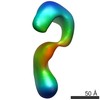
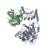
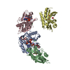
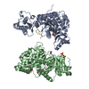
| Title | Crystal structure of Saccharomyces cerevisiae NUP192, residues 2 to 960 [ScNup192(2-960)] | ||||||
 Components Components | Nucleoporin NUP192 | ||||||
 Keywords Keywords | PROTEIN TRANSPORT / Structural genomics / NYSGRC / PSI-Biology / New York Structural Genomics Research Consortium / alpha solenoid-like / Nuclear Pore Complex component / NPC / Nup192 / Nup188 / Nucleoporin / Nucleocytoplasmic Transport: a Target for Cellular Control / NPCXstals | ||||||
| Function / homology |  Function and homology information Function and homology informationnuclear pore inner ring / regulation of nucleocytoplasmic transport / nuclear pore organization / structural constituent of nuclear pore / nucleocytoplasmic transport / nuclear pore / mRNA transport / nuclear envelope / protein transport / nucleus Similarity search - Function | ||||||
| Biological species |  | ||||||
| Method |  X-RAY DIFFRACTION / SYNCHROTRON / SAD / Resolution: 3.25 Å X-RAY DIFFRACTION / SYNCHROTRON / SAD / Resolution: 3.25 Å | ||||||
 Authors Authors | Sampathkumar, P. / Almo, S.C. / New York Structural Genomics Research Consortium (NYSGRC) / Nucleocytoplasmic Transport: a Target for Cellular Control (NPCXstals) | ||||||
 Citation Citation | Journal: Structure / Year: 2013 Title: Structure, dynamics, evolution, and function of a major scaffold component in the nuclear pore complex. Authors: Parthasarathy Sampathkumar / Seung Joong Kim / Paula Upla / William J Rice / Jeremy Phillips / Benjamin L Timney / Ursula Pieper / Jeffrey B Bonanno / Javier Fernandez-Martinez / Zhanna ...Authors: Parthasarathy Sampathkumar / Seung Joong Kim / Paula Upla / William J Rice / Jeremy Phillips / Benjamin L Timney / Ursula Pieper / Jeffrey B Bonanno / Javier Fernandez-Martinez / Zhanna Hakhverdyan / Natalia E Ketaren / Tsutomu Matsui / Thomas M Weiss / David L Stokes / J Michael Sauder / Stephen K Burley / Andrej Sali / Michael P Rout / Steven C Almo /  Abstract: The nuclear pore complex, composed of proteins termed nucleoporins (Nups), is responsible for nucleocytoplasmic transport in eukaryotes. Nuclear pore complexes (NPCs) form an annular structure ...The nuclear pore complex, composed of proteins termed nucleoporins (Nups), is responsible for nucleocytoplasmic transport in eukaryotes. Nuclear pore complexes (NPCs) form an annular structure composed of the nuclear ring, cytoplasmic ring, a membrane ring, and two inner rings. Nup192 is a major component of the NPC's inner ring. We report the crystal structure of Saccharomyces cerevisiae Nup192 residues 2-960 [ScNup192(2-960)], which adopts an α-helical fold with three domains (i.e., D1, D2, and D3). Small angle X-ray scattering and electron microscopy (EM) studies reveal that ScNup192(2-960) could undergo long-range transition between "open" and "closed" conformations. We obtained a structural model of full-length ScNup192 based on EM, the structure of ScNup192(2-960), and homology modeling. Evolutionary analyses using the ScNup192(2-960) structure suggest that NPCs and vesicle-coating complexes are descended from a common membrane-coating ancestral complex. We show that suppression of Nup192 expression leads to compromised nuclear transport and hypothesize a role for Nup192 in modulating the permeability of the NPC central channel. | ||||||
| History |
|
- Structure visualization
| Structure viewer | Molecule: MolmilJmol/JSmol |
|---|
- Downloads & links
-Download
| PDBx/mmCIF format | 4ifq.cif.gz | 188.9 KB | Display | PDBx/mmCIF format |
|---|---|---|---|---|
| PDB format | pdb4ifq.ent.gz | 147.8 KB | Display | PDB format |
| PDBx/mmJSON format | 4ifq.json.gz | Tree view | PDBx/mmJSON format | |
| Others |  Other downloads Other downloads |
-Validation report
| Arichive directory | https://data.pdbj.org/pub/pdb/validation_reports/if/4ifqftp://data.pdbj.org/pub/pdb/validation_reports/if/4ifq | HTTPS FTP |
|---|
-Related structure data


-Links
 PDBj
PDBj
- Assembly
| Deposited unit | 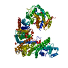
| ||||||||
|---|---|---|---|---|---|---|---|---|---|
| 1 |
| ||||||||
| 2 | 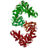
| ||||||||
| Unit cell |
| ||||||||
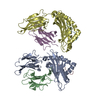
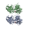
| Details | THE DIMERIC INTERFACE SUGGESTED BY PISA IS LIKELY TO BE NON-BIOLOGICAL |
-Components
| #1: Protein | Mass: 111779.430 Da / Num. of mol.: 1 / Fragment: UNP residues 2-960 Source method: isolated from a genetically manipulated source Source: (gene. exp.) Strain: ATCC 204508 / S288c / Gene: J1216, NUP192, YJL039C / Plasmid: pSGX3 / Production host:  | ||||||
|---|---|---|---|---|---|---|---|
| #2: Chemical | ChemComp-SO4 /   Mass: 96.063 Da / Num. of mol.: 7 / Source method: obtained synthetically / Formula: SO4 Mass: 96.063 Da / Num. of mol.: 7 / Source method: obtained synthetically / Formula: SO4#3: Chemical | ChemComp-IOD /   Mass: 126.904 Da / Num. of mol.: 7 / Source method: obtained synthetically / Formula: I Mass: 126.904 Da / Num. of mol.: 7 / Source method: obtained synthetically / Formula: I#4: Water | ChemComp-HOH / |  Mass: 18.015 Da / Num. of mol.: 6 / Source method: isolated from a natural source / Formula: H2O Mass: 18.015 Da / Num. of mol.: 6 / Source method: isolated from a natural source / Formula: H2OHas protein modification | Y | |
-Experimental details
-Experiment
| Experiment | Method: X-RAY DIFFRACTION / Number of used crystals: 1 |
|---|
- Sample preparation
| Crystal | Density Matthews: 4.76 Å3/Da / Density % sol: 74.14 % |
|---|---|
| Crystal grow | Temperature: 298 K / Method: vapor diffusion, sitting drop Details: Protein (20 mM Hepes, pH 8.0, 500 mM NaCl, 10% glycerol, 5mM DTT; Reservoir (10% PEG3350, 100mM pottasium iodide); Cryoprotection (30% PEG400 and 25% saturated ammonium sulfate), Vapor ...Details: Protein (20 mM Hepes, pH 8.0, 500 mM NaCl, 10% glycerol, 5mM DTT; Reservoir (10% PEG3350, 100mM pottasium iodide); Cryoprotection (30% PEG400 and 25% saturated ammonium sulfate), Vapor Diffusion, Sitting Drop, temperature 298K |
-Data collection
| Diffraction | Mean temperature: 100 K |
|---|---|
| Diffraction source | Source: SYNCHROTRON / Site: NSLS / Beamline: X29A / Wavelength: 0.9792 Å |
| Detector | Type: ADSC QUANTUM 315 / Detector: CCD / Date: Mar 15, 2012 / Details: MIRRORS |
| Radiation | Monochromator: Si(111) / Protocol: SINGLE WAVELENGTH / Monochromatic (M) / Laue (L): M / Scattering type: x-ray |
| Radiation wavelength | Wavelength: 0.9792 Å / Relative weight: 1 |
| Reflection | Resolution: 3.254→50 Å / Num. all: 64698 / Num. obs: 64698 / % possible obs: 100 % / Observed criterion σ(I): 0 / Redundancy: 8.4 % / Biso Wilson estimate: 78 Å2 / Rsym value: 0.136 / Net I/σ(I): 17.5 |
| Reflection shell | Resolution: 3.25→3.29 Å / Redundancy: 8.4 % / Mean I/σ(I) obs: 2.6 / Num. unique all: 2186 / Rsym value: 0.969 / % possible all: 100 |
- Processing
| Software |
| ||||||||||||||||||||||||||||||||||||||||||||||||||||||||||||
|---|---|---|---|---|---|---|---|---|---|---|---|---|---|---|---|---|---|---|---|---|---|---|---|---|---|---|---|---|---|---|---|---|---|---|---|---|---|---|---|---|---|---|---|---|---|---|---|---|---|---|---|---|---|---|---|---|---|---|---|---|---|
| Refinement | Method to determine structure: SAD Starting model: Built using AutoBuild (Phenix) and Buccaneer (CCP4) Resolution: 3.25→47.76 Å / Cor.coef. Fo:Fc: 0.944 / Cor.coef. Fo:Fc free: 0.91 / Occupancy max: 1 / Occupancy min: 0.45 / SU B: 15.519 / SU ML: 0.253 / Cross valid method: THROUGHOUT / σ(F): 0 / ESU R: 0.753 / ESU R Free: 0.373 / Stereochemistry target values: MAXIMUM LIKELIHOOD Details: HYDROGENS HAVE BEEN ADDED IN THE RIDING POSITIONS U VALUES : REFINED INDIVIDUALLY
| ||||||||||||||||||||||||||||||||||||||||||||||||||||||||||||
| Solvent computation | Ion probe radii: 0.8 Å / Shrinkage radii: 0.8 Å / VDW probe radii: 1.2 Å / Solvent model: BABINET MODEL WITH MASK | ||||||||||||||||||||||||||||||||||||||||||||||||||||||||||||
| Displacement parameters | Biso max: 184.54 Å2 / Biso mean: 85.2254 Å2 / Biso min: 41.66 Å2
| ||||||||||||||||||||||||||||||||||||||||||||||||||||||||||||
| Refinement step | Cycle: LAST / Resolution: 3.25→47.76 Å
| ||||||||||||||||||||||||||||||||||||||||||||||||||||||||||||
| Refine LS restraints |
| ||||||||||||||||||||||||||||||||||||||||||||||||||||||||||||
| LS refinement shell | Resolution: 3.254→3.338 Å / Total num. of bins used: 20
|
