 Movie
Movie Controller


+ Open data

- Basic information
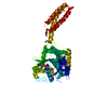
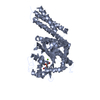
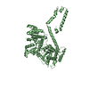
| Entry | Database: PDB / ID: 4idp | ||||||
|---|---|---|---|---|---|---|---|
| Title | human atlastin-1 1-446, N440T, GppNHp | ||||||
 Components Components | Atlastin-1 | ||||||
 Keywords Keywords | HYDROLASE / GTPase / GTP/GDP binding | ||||||
| Function / homology |  Function and homology information Function and homology informationendoplasmic reticulum tubular network membrane / endoplasmic reticulum tubular network membrane organization / Golgi cis cisterna membrane / GTPase-dependent fusogenic activity / endoplasmic reticulum tubular network / endoplasmic reticulum membrane fusion / endoplasmic reticulum organization / axonogenesis / protein homooligomerization / Hydrolases; Acting on acid anhydrides; Acting on GTP to facilitate cellular and subcellular movement ...endoplasmic reticulum tubular network membrane / endoplasmic reticulum tubular network membrane organization / Golgi cis cisterna membrane / GTPase-dependent fusogenic activity / endoplasmic reticulum tubular network / endoplasmic reticulum membrane fusion / endoplasmic reticulum organization / axonogenesis / protein homooligomerization / Hydrolases; Acting on acid anhydrides; Acting on GTP to facilitate cellular and subcellular movement / Golgi membrane / axon / GTPase activity / endoplasmic reticulum membrane / GTP binding / Golgi apparatus / endoplasmic reticulum / identical protein binding Similarity search - Function | ||||||
| Biological species |  Homo sapiens (human) Homo sapiens (human) | ||||||
| Method |  X-RAY DIFFRACTION / SYNCHROTRON / MOLECULAR REPLACEMENT / Resolution: 2.587 Å X-RAY DIFFRACTION / SYNCHROTRON / MOLECULAR REPLACEMENT / Resolution: 2.587 Å | ||||||
 Authors Authors | Byrnes, L.J. / Singh, A. / Szeto, K. / Benvin, N.M. / O'Donnell, J.P. / Zipfel, W.R. / Sondermann, H. | ||||||
 Citation Citation | Journal: Embo J. / Year: 2013 Title: Structural basis for conformational switching and GTP loading of the large G protein atlastin. Authors: Byrnes, L.J. / Singh, A. / Szeto, K. / Benvin, N.M. / O'Donnell, J.P. / Zipfel, W.R. / Sondermann, H. | ||||||
| History |
|
- Structure visualization
| Structure viewer | Molecule: MolmilJmol/JSmol |
|---|
- Downloads & links
-Download
| PDBx/mmCIF format | 4idp.cif.gz | 668.3 KB | Display | PDBx/mmCIF format |
|---|---|---|---|---|
| PDB format | pdb4idp.ent.gz | 554.9 KB | Display | PDB format |
| PDBx/mmJSON format | 4idp.json.gz | Tree view | PDBx/mmJSON format | |
| Others |  Other downloads Other downloads |
-Validation report
| Arichive directory | https://data.pdbj.org/pub/pdb/validation_reports/id/4idpftp://data.pdbj.org/pub/pdb/validation_reports/id/4idp | HTTPS FTP |
|---|
-Related structure data
| Related structure data | 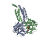 4idnC 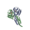 4idoC 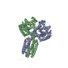 4idqSC C: citing same article ( S: Starting model for refinement |
|---|---|
| Similar structure data |


-Links
 PDBj
PDBj


- Assembly
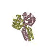
| Deposited unit | 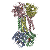
| ||||||||
|---|---|---|---|---|---|---|---|---|---|
| 1 | 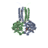
| ||||||||
| 2 | 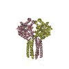
| ||||||||
| Unit cell |
|
-Components
| #1: Protein | Mass: 51760.031 Da / Num. of mol.: 4 / Fragment: cytoplasmic domain (UNP residues 1-446) / Mutation: N440T Source method: isolated from a genetically manipulated source Source: (gene. exp.) Homo sapiens (human) / Gene: ATL1, GBP3, SPG3A / Plasmid: pET28 / Production host:  References: UniProt: Q8WXF7, Hydrolases; Acting on acid anhydrides; Acting on GTP to facilitate cellular and subcellular movement #2: Chemical | ChemComp-GNP /   Mass: 522.196 Da / Num. of mol.: 4 / Source method: obtained synthetically / Formula: C10H17N6O13P3 Mass: 522.196 Da / Num. of mol.: 4 / Source method: obtained synthetically / Formula: C10H17N6O13P3Comment: GppNHp, GMPPNP, energy-carrying molecule analogue*YM #3: Chemical | ChemComp-MG /   Mass: 24.305 Da / Num. of mol.: 4 / Source method: obtained synthetically / Formula: Mg Mass: 24.305 Da / Num. of mol.: 4 / Source method: obtained synthetically / Formula: Mg#4: Water | ChemComp-HOH / |  Mass: 18.015 Da / Num. of mol.: 403 / Source method: isolated from a natural source / Formula: H2O Mass: 18.015 Da / Num. of mol.: 403 / Source method: isolated from a natural source / Formula: H2OHas protein modification | Y | |
|---|
-Experimental details
-Experiment
| Experiment | Method: X-RAY DIFFRACTION / Number of used crystals: 1 |
|---|
- Sample preparation
| Crystal | Density Matthews: 2.67 Å3/Da / Density % sol: 53.88 % |
|---|---|
| Crystal grow | Temperature: 293 K / Method: vapor diffusion, hanging drop / pH: 8.4 Details: 0.2 M lithium citrate tribasic tetrahydrate, 20% PEG3350, pH 8.4, VAPOR DIFFUSION, HANGING DROP, temperature 293K |
-Data collection
| Diffraction | Mean temperature: 100 K |
|---|---|
| Diffraction source | Source: SYNCHROTRON / Site: CHESS  / Beamline: A1 / Wavelength: 0.987 Å / Beamline: A1 / Wavelength: 0.987 Å |
| Detector | Type: ADSC QUANTUM 210 / Detector: CCD / Date: May 4, 2011 |
| Radiation | Monochromator: Si(111) / Protocol: SINGLE WAVELENGTH / Monochromatic (M) / Laue (L): M / Scattering type: x-ray |
| Radiation wavelength | Wavelength: 0.987 Å / Relative weight: 1 |
| Reflection | Resolution: 2.587→50 Å / Num. all: 69849 / Num. obs: 69639 / % possible obs: 99.7 % / Observed criterion σ(F): 1 / Observed criterion σ(I): 1 / Redundancy: 7.8 % / Rsym value: 0.16 / Net I/σ(I): 12.3 |
| Reflection shell | Resolution: 2.587→2.69 Å / Redundancy: 6.7 % / Mean I/σ(I) obs: 3.1 / Rsym value: 0.611 / % possible all: 97.4 |
- Processing
| Software |
| |||||||||||||||||||||||||||||||||||||||||||||||||||||||||||||||||||||||||||||||||||||||||||||||||||||||||||||||||||||||||||||||||||||||||||||||||||||||||||||||||||||||||||||||||||||||||||||||||||||||||||||||||||||||||||||||||
|---|---|---|---|---|---|---|---|---|---|---|---|---|---|---|---|---|---|---|---|---|---|---|---|---|---|---|---|---|---|---|---|---|---|---|---|---|---|---|---|---|---|---|---|---|---|---|---|---|---|---|---|---|---|---|---|---|---|---|---|---|---|---|---|---|---|---|---|---|---|---|---|---|---|---|---|---|---|---|---|---|---|---|---|---|---|---|---|---|---|---|---|---|---|---|---|---|---|---|---|---|---|---|---|---|---|---|---|---|---|---|---|---|---|---|---|---|---|---|---|---|---|---|---|---|---|---|---|---|---|---|---|---|---|---|---|---|---|---|---|---|---|---|---|---|---|---|---|---|---|---|---|---|---|---|---|---|---|---|---|---|---|---|---|---|---|---|---|---|---|---|---|---|---|---|---|---|---|---|---|---|---|---|---|---|---|---|---|---|---|---|---|---|---|---|---|---|---|---|---|---|---|---|---|---|---|---|---|---|---|---|---|---|---|---|---|---|---|---|---|---|---|---|---|---|---|---|
| Refinement | Method to determine structure: MOLECULAR REPLACEMENT Starting model: PDB ENTRY 4IDQ Resolution: 2.587→49.678 Å / SU ML: 0.29 / σ(F): 0.32 / Phase error: 32.25 / Stereochemistry target values: MLHL
| |||||||||||||||||||||||||||||||||||||||||||||||||||||||||||||||||||||||||||||||||||||||||||||||||||||||||||||||||||||||||||||||||||||||||||||||||||||||||||||||||||||||||||||||||||||||||||||||||||||||||||||||||||||||||||||||||
| Solvent computation | Shrinkage radii: 0.9 Å / VDW probe radii: 1.11 Å / Solvent model: FLAT BULK SOLVENT MODEL | |||||||||||||||||||||||||||||||||||||||||||||||||||||||||||||||||||||||||||||||||||||||||||||||||||||||||||||||||||||||||||||||||||||||||||||||||||||||||||||||||||||||||||||||||||||||||||||||||||||||||||||||||||||||||||||||||
| Refinement step | Cycle: LAST / Resolution: 2.587→49.678 Å
| |||||||||||||||||||||||||||||||||||||||||||||||||||||||||||||||||||||||||||||||||||||||||||||||||||||||||||||||||||||||||||||||||||||||||||||||||||||||||||||||||||||||||||||||||||||||||||||||||||||||||||||||||||||||||||||||||
| Refine LS restraints |
| |||||||||||||||||||||||||||||||||||||||||||||||||||||||||||||||||||||||||||||||||||||||||||||||||||||||||||||||||||||||||||||||||||||||||||||||||||||||||||||||||||||||||||||||||||||||||||||||||||||||||||||||||||||||||||||||||
| LS refinement shell |
| |||||||||||||||||||||||||||||||||||||||||||||||||||||||||||||||||||||||||||||||||||||||||||||||||||||||||||||||||||||||||||||||||||||||||||||||||||||||||||||||||||||||||||||||||||||||||||||||||||||||||||||||||||||||||||||||||
| Refinement TLS params. | Method: refined / Refine-ID: X-RAY DIFFRACTION
| |||||||||||||||||||||||||||||||||||||||||||||||||||||||||||||||||||||||||||||||||||||||||||||||||||||||||||||||||||||||||||||||||||||||||||||||||||||||||||||||||||||||||||||||||||||||||||||||||||||||||||||||||||||||||||||||||
| Refinement TLS group |
|
