- PDB-4iaj: Crystal structure of a conserved domain protein (SP_1775) from St... -
+
Open data
ID or keywords:
Loading...
-
Basic information
Entry
Database: PDB / ID: 4iaj
Title
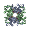
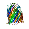
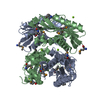
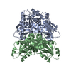
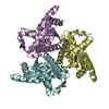
Crystal structure of a conserved domain protein (SP_1775) from Streptococcus pneumoniae TIGR4 at 1.91 A resolution
Components
Conserved domain protein
Keywords
Structural Genomics / Unknown Function / DUF4649 / PF15507 family protein / Joint Center for Structural Genomics / JCSG / Protein Structure Initiative / PSI-BIOLOGY
Function / homology
Protein of unknown function DUF4649 / Protein of unknown function DUF4649 / Domain of unknown function (DUF4649) / Dna Ligase; domain 1 / 2-Layer Sandwich / Alpha Beta / Unknown ligand / Conserved domain protein / Conserved domain protein
Function and homology information
Biological species
Streptococcus pneumoniae (bacteria)
Method
X-RAY DIFFRACTION / SYNCHROTRON / MAD / Resolution: 1.91 Å
#191 - Nov 2015 Glutamate-gated Chloride Receptors similarity (1)
-
Assembly
Deposited unit
A: Conserved domain protein B: Conserved domain protein C: Conserved domain protein D: Conserved domain protein E: Conserved domain protein F: Conserved domain protein G: Conserved domain protein H: Conserved domain protein hetero molecules
Mass: 18.015 Da / Num. of mol.: 309 / Source method: isolated from a natural source / Formula: H2O
-
Details
Has protein modification
Y
Sequence details
THIS CONSTRUCT (RESIDUES 24-100) WAS EXPRESSED WITH AN N-TERMINAL PURIFICATION TAG ...THIS CONSTRUCT (RESIDUES 24-100) WAS EXPRESSED WITH AN N-TERMINAL PURIFICATION TAG MGSDKIHHHHHHENLYFQG. THE TAG WAS REMOVED WITH TEV PROTEASE LEAVING ONLY A GLYCINE (0) FOLLOWED BY THE TARGET SEQUENCE.
-
Experimental details
-
Experiment
Experiment
Method: X-RAY DIFFRACTION / Number of used crystals: 1
-
Sample preparation
Crystal
Density Matthews: 2.2 Å3/Da / Density % sol: 44.01 %
Crystal grow
Temperature: 277 K / Method: vapor diffusion, sitting drop Details: 0.2M magnesium nitrate, 20.0% polyethylene glycol 3350, NANODROP, VAPOR DIFFUSION, SITTING DROP, temperature 277K
Resolution: 1.91→29.304 Å / Num. obs: 48771 / % possible obs: 96.2 % / Observed criterion σ(I): -3 / Biso Wilson estimate: 26.14 Å2 / Rmerge(I) obs: 0.051 / Net I/σ(I): 10.68
Reflection shell
Diffraction-ID: 1
Resolution (Å)
Highest resolution (Å)
Rmerge(I) obs
Mean I/σ(I) obs
Num. measured obs
Num. unique obs
% possible all
1.91-1.98
0.564
1.5
15463
9493
95.4
1.98-2.06
0.366
2.1
15407
9448
95.9
2.06-2.15
0.277
2.7
14550
8938
95.8
2.15-2.26
0.208
3.8
14958
9207
96.3
2.26-2.41
0.151
5.1
16224
9980
96.8
2.41-2.59
0.1
7.4
14750
9080
96.6
2.59-2.85
0.069
9.9
15379
9500
97.1
2.85-3.26
0.04
16.1
15160
9382
96.4
3.26-4.1
0.023
25.5
14899
9321
95.4
4.1
0.017
32.4
15278
9504
95.9
-
Phasing
Phasing
Method: MAD
-
Processing
Software
Name
Version
Classification
NB
MolProbity
3beta29
modelbuilding
PDB_EXTRACT
3.1
dataextraction
SHELX
phasing
SHARP
phasing
XSCALE
March15, 2012
datascaling
REFMAC
5.7.0032
refinement
XDS
datareduction
SHELXD
phasing
Refinement
Method to determine structure: MAD / Resolution: 1.91→29.304 Å / Cor.coef. Fo:Fc: 0.96 / Cor.coef. Fo:Fc free: 0.934 / Occupancy max: 1 / Occupancy min: 0.33 / SU B: 7.466 / SU ML: 0.111 / Cross valid method: THROUGHOUT / σ(F): 0 / ESU R: 0.171 / ESU R Free: 0.158 Stereochemistry target values: MAXIMUM LIKELIHOOD WITH PHASES Details: 1.HYDROGENS HAVE BEEN ADDED IN THE RIDING POSITIONS. 2.A MET-INHIBITION PROTOCOL WAS USED FOR SELENOMETHIONINE INCORPORATION DURING PROTEIN EXPRESSION. THE OCCUPANCY OF THE SE ATOMS IN THE ...Details: 1.HYDROGENS HAVE BEEN ADDED IN THE RIDING POSITIONS. 2.A MET-INHIBITION PROTOCOL WAS USED FOR SELENOMETHIONINE INCORPORATION DURING PROTEIN EXPRESSION. THE OCCUPANCY OF THE SE ATOMS IN THE MSE RESIDUES WAS REDUCED TO 0.75 FOR THE REDUCED SCATTERING POWER DUE TO PARTIAL S-MET INCORPORATION. 3.ATOM RECORDS CONTAIN SUM OF TLS AND RESIDUAL B FACTORS. ANISOU RECORD CONTAINS SUM OF TLS AND RESIDUAL U FACTORS. 4.WATERS WERE EXCLUDED FROM AUTOMATIC TLS ASSIGNMENT. 5.MAGNESIUM (MG) AND 1,2-ETHANEDIOL FROM THE CRYSTALLIZATION/CRYO CONDITIONS HAVE BEEN MODELED INTO THE STRUCTURE. 6. UNKNOWN LIGANDS (UNL) AND UNKNOWN ATOMS (UNX) HAVE BEEN MODELED INTO DIFFERENCE ELECTRON DENSITY WITHIN A CHANNEL FORMED BY THE OCTAMERIC ASSEMBLY IN THE ASYMMETRIC UNIT.
Rfactor
Num. reflection
% reflection
Selection details
Rfree
0.2324
2471
5.1 %
RANDOM
Rwork
0.1798
-
-
-
obs
0.1824
48740
98.58 %
-
Solvent computation
Ion probe radii: 0.8 Å / Shrinkage radii: 0.8 Å / VDW probe radii: 1.2 Å / Solvent model: BABINET MODEL WITH MASK
 Movie
Movie Controller

 Yorodumi
 Open data
 Basic information
 Components
 Keywords
 Function and homology information
 Streptococcus pneumoniae (bacteria)
 X-RAY DIFFRACTION /
  Authors
 Citation
 Structure visualization
 Downloads & links
 Other downloads
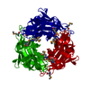
 PDBj
PDBj

 Assembly
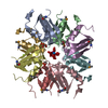


 Num. of mol.: 5 / Source method: obtained synthetically
 Mass: 62.068 Da / Num. of mol.: 8 / Source method: obtained synthetically / Formula: C2H6O2
 Mass: 24.305 Da / Num. of mol.: 3 / Source method: obtained synthetically / Formula: Mg
 Sample preparation
 / Beamline: 8.2.2 / Wavelength: 0.918401,0.979338,0.979111
 Processing