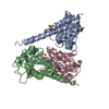
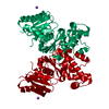
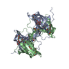
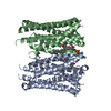
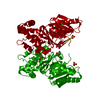
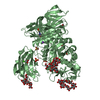
- PDB-4i95: Crystal structure of a Lipocalin-like protein (BACEGG_00036) from... -
+
Open data
ID or keywords:
Loading...
-
Basic information
Entry
Database: PDB / ID: 4i95
Title
Crystal structure of a Lipocalin-like protein (BACEGG_00036) from Bacteroides eggerthii DSM 20697 at 1.81 A resolution
Components
Putative uncharacterized protein
Keywords
STRUCTURAL GENOMICS / UNKNOWN FUNCTION / Lipocalin-like domain of PF13924 family / Joint Center for Structural Genomics / JCSG / Protein Structure Initiative / PSI-BIOLOGY
Mass: 18.015 Da / Num. of mol.: 293 / Source method: isolated from a natural source / Formula: H2O
Has protein modification
Y
Sequence details
THIS CONSTRUCT (RESIDUES 23-163) WAS EXPRESSED WITH A PURIFICATION TAG MGSDKIHHHHHHENLYFQG. THE TAG ...THIS CONSTRUCT (RESIDUES 23-163) WAS EXPRESSED WITH A PURIFICATION TAG MGSDKIHHHHHHENLYFQG. THE TAG WAS REMOVED WITH TEV PROTEASE LEAVING ONLY A GLYCINE (0) FOLLOWED BY THE TARGET SEQUENCE.
-
Experimental details
-
Experiment
Experiment
Method: X-RAY DIFFRACTION / Number of used crystals: 1
-
Sample preparation
Crystal
Density Matthews: 2.69 Å3/Da / Density % sol: 54.32 %
Crystal grow
Temperature: 277 K / Method: vapor diffusion, sitting drop / pH: 6 Details: 1.0000M lithium chloride, 20.0000% polyethylene glycol 6000, 0.1M MES pH 6.0, NANODROP, VAPOR DIFFUSION, SITTING DROP, temperature 277K
Monochromator: double crystal Si(111) / Protocol: SINGLE WAVELENGTH / Monochromatic (M) / Laue (L): M / Scattering type: x-ray
Radiation wavelength
Wavelength: 0.9795 Å / Relative weight: 1
Reflection
Resolution: 1.81→48.331 Å / Num. obs: 33477 / % possible obs: 100 % / Observed criterion σ(I): -3 / Biso Wilson estimate: 21.921 Å2 / Rmerge(I) obs: 0.193 / Net I/σ(I): 15.74
Reflection shell
Rmerge(I) obs: 0.016 / Diffraction-ID: 1
Resolution (Å)
Highest resolution (Å)
Mean I/σ(I) obs
Num. measured obs
Num. unique obs
% possible all
1.81-1.87
2.9
123609
3053
100
1.87-1.95
4.5
140410
3515
100
1.95-2.04
6
128102
3311
100
2.04-2.15
7.8
130915
3369
100
2.15-2.28
10.4
128077
3165
100
2.28-2.46
12.7
131880
3393
100
2.46-2.7
15.3
126392
3238
100
2.7-3.09
22.1
133569
3363
99.9
3.09-3.89
33.3
131836
3417
100
3.89
39
132442
3653
99.8
-
Phasing
Phasing
Method: SAD
-
Processing
Software
Name
Version
Classification
NB
MolProbity
3beta29
modelbuilding
PDB_EXTRACT
3.1
dataextraction
SHELX
phasing
SHARP
phasing
XSCALE
March15, 2012
datascaling
REFMAC
5.7.0032
refinement
XDS
datareduction
SHELXD
phasing
Refinement
Method to determine structure: SAD / Resolution: 1.81→48.331 Å / Cor.coef. Fo:Fc: 0.969 / Cor.coef. Fo:Fc free: 0.962 / Occupancy max: 1 / Occupancy min: 0.3 / SU B: 4.392 / SU ML: 0.07 / Cross valid method: THROUGHOUT / σ(F): 0 / ESU R: 0.111 / ESU R Free: 0.107 Stereochemistry target values: MAXIMUM LIKELIHOOD WITH PHASES Details: 1.HYDROGENS HAVE BEEN ADDED IN THE RIDING POSITIONS. 2.A MET-INHIBITION PROTOCOL WAS USED FOR SELENOMETHIONINE INCORPORATION DURING PROTEIN EXPRESSION. THE OCCUPANCY OF THE SE ATOMS IN THE ...Details: 1.HYDROGENS HAVE BEEN ADDED IN THE RIDING POSITIONS. 2.A MET-INHIBITION PROTOCOL WAS USED FOR SELENOMETHIONINE INCORPORATION DURING PROTEIN EXPRESSION. THE OCCUPANCY OF THE SE ATOMS IN THE MSE RESIDUES WAS REDUCED TO 0.75 FOR THE REDUCED SCATTERING POWER DUE TO PARTIAL S-MET INCORPORATION. 3.ATOM RECORDS CONTAIN SUM OF TLS AND RESIDUAL B FACTORS. ANISOU RECORD CONTAINS SUM OF TLS AND RESIDUAL U FACTORS. 4.WATERS WERE EXCLUDED FROM AUTOMATIC TLS ASSIGNMENT. 5.AN UNIDENTIFIED LIGAND (UNL) HAS BEEN MODELED AT THE PUTATIVE ACTIVE SITE IN BOTH MONOMERS.
Rfactor
Num. reflection
% reflection
Selection details
Rfree
0.2039
1693
5.1 %
RANDOM
Rwork
0.1738
-
-
-
obs
0.1753
33450
99.98 %
-
Solvent computation
Ion probe radii: 0.8 Å / Shrinkage radii: 0.8 Å / VDW probe radii: 1.2 Å / Solvent model: BABINET MODEL WITH MASK
In the structure databanks used in Yorodumi, some data are registered as the other names, "COVID-19 virus" and "2019-nCoV". Here are the details of the virus and the list of structure data.
Jan 31, 2019. EMDB accession codes are about to change! (news from PDBe EMDB page)
EMDB accession codes are about to change! (news from PDBe EMDB page)
The allocation of 4 digits for EMDB accession codes will soon come to an end. Whilst these codes will remain in use, new EMDB accession codes will include an additional digit and will expand incrementally as the available range of codes is exhausted. The current 4-digit format prefixed with “EMD-” (i.e. EMD-XXXX) will advance to a 5-digit format (i.e. EMD-XXXXX), and so on. It is currently estimated that the 4-digit codes will be depleted around Spring 2019, at which point the 5-digit format will come into force.
The EM Navigator/Yorodumi systems omit the EMD- prefix.
Related info.:Q: What is EMD? / ID/Accession-code notation in Yorodumi/EM Navigator
Yorodumi is a browser for structure data from EMDB, PDB, SASBDB, etc.
This page is also the successor to EM Navigator detail page, and also detail information page/front-end page for Omokage search.
The word "yorodu" (or yorozu) is an old Japanese word meaning "ten thousand". "mi" (miru) is to see.
Related info.:EMDB / PDB / SASBDB / Comparison of 3 databanks / Yorodumi Search / Aug 31, 2016. New EM Navigator & Yorodumi / Yorodumi Papers / Jmol/JSmol / Function and homology information / Changes in new EM Navigator and Yorodumi
 Movie
Movie Controller

 Yorodumi
 Open data
 Basic information
 Components
 Keywords
 Function and homology information
 Bacteroides eggerthii (bacteria)
 X-RAY DIFFRACTION /
  Authors
 Citation
 Structure visualization
 Downloads & links
 Other downloads
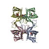

 PDBj
PDBj

 Assembly

 Mass: 18.015 Da / Num. of mol.: 293 / Source method: isolated from a natural source / Formula: H2O
 Sample preparation
 / Beamline: BL12-2 / Wavelength: 0.9795
  Processing