 Movie
Movie Controller

[English] 日本語
 Yorodumi
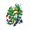
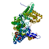
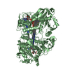
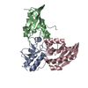
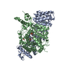
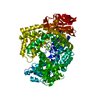
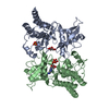
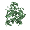
- PDB-4hzb: Crystal structure of the type VI SeMet effector-immunity complex ... -
+ Open data

- Basic information
| Entry | Database: PDB / ID: 4hzb | ||||||
|---|---|---|---|---|---|---|---|
| Title | Crystal structure of the type VI SeMet effector-immunity complex Tae3-Tai3 from Ralstonia pickettii | ||||||
 Components Components |
| ||||||
 Keywords Keywords | HYDROLASE / protein-protein complex / alpha+beta / endopeptidase / periplasmic space | ||||||
| Function / homology |  Function and homology information Function and homology informationDomain of unknown function DUF3828 / Protein of unknown function (DUF3828) / endopeptidase domain like (from Nostoc punctiforme) / endopeptidase fold (from Nostoc punctiforme) / Nuclear Transport Factor 2; Chain: A, - #50 / Nuclear Transport Factor 2; Chain: A, / Roll / Alpha-Beta Complex / Alpha Beta Similarity search - Domain/homology | ||||||
| Biological species |  Ralstonia pickettii (bacteria) Ralstonia pickettii (bacteria) | ||||||
| Method |  X-RAY DIFFRACTION / SYNCHROTRON / SAD / Resolution: 2.6 Å X-RAY DIFFRACTION / SYNCHROTRON / SAD / Resolution: 2.6 Å | ||||||
 Authors Authors | Dong, C. / Zhang, H. / Gao, Z.Q. / Dong, Y.H. | ||||||
 Citation Citation | Journal: Biochem.J. / Year: 2013 Title: Structural insights into the inhibition of type VI effector Tae3 by its immunity protein Tai3 Authors: Dong, C. / Zhang, H. / Gao, Z.Q. / Wang, W.J. / She, Z. / Liu, G.F. / Shen, Y.Q. / Su, X.D. / Dong, Y.H. | ||||||
| History |
|
- Structure visualization
| Structure viewer | Molecule: MolmilJmol/JSmol |
|---|
- Downloads & links
-Download
| PDBx/mmCIF format | 4hzb.cif.gz | 153.3 KB | Display | PDBx/mmCIF format |
|---|---|---|---|---|
| PDB format | pdb4hzb.ent.gz | 122.6 KB | Display | PDB format |
| PDBx/mmJSON format | 4hzb.json.gz | Tree view | PDBx/mmJSON format | |
| Others |  Other downloads Other downloads |
-Validation report
| Arichive directory | https://data.pdbj.org/pub/pdb/validation_reports/hz/4hzbftp://data.pdbj.org/pub/pdb/validation_reports/hz/4hzb | HTTPS FTP |
|---|
-Related structure data


-Links
 PDBj
PDBj- Assembly
| Deposited unit | 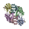
| ||||||||
|---|---|---|---|---|---|---|---|---|---|
| 1 |
| ||||||||
| Unit cell |
|
-Components
| #1: Protein | Mass: 13241.492 Da / Num. of mol.: 2 Source method: isolated from a genetically manipulated source Source: (gene. exp.) Ralstonia pickettii (bacteria) / Strain: 12D / Gene: Rpic12D_3261 / Production host: #2: Protein | Mass: 17311.689 Da / Num. of mol.: 4 Source method: isolated from a genetically manipulated source Source: (gene. exp.) Ralstonia pickettii (bacteria) / Strain: 12D / Gene: Rpic12D_3260 / Production host: #3: Water | ChemComp-HOH / |  Mass: 18.015 Da / Num. of mol.: 161 / Source method: isolated from a natural source / Formula: H2O Mass: 18.015 Da / Num. of mol.: 161 / Source method: isolated from a natural source / Formula: H2OHas protein modification | Y | |
|---|
-Experimental details
-Experiment
| Experiment | Method: X-RAY DIFFRACTION / Number of used crystals: 1 |
|---|
- Sample preparation
| Crystal | Density Matthews: 2.7 Å3/Da / Density % sol: 54.5 % |
|---|---|
| Crystal grow | Temperature: 277 K / Method: vapor diffusion, sitting drop / pH: 5.6 Details: 0.01M ferric chloride hexahydrate, 0.1M tri-sodium citrate dihydrate pH5.6, 10% v/v jeffamine M-600 , pH 5.6 , VAPOR DIFFUSION, SITTING DROP, temperature 277 K |
-Data collection
| Diffraction | Mean temperature: 100 K |
|---|---|
| Diffraction source | Source: SYNCHROTRON / Site: BSRF  / Beamline: 3W1A / Wavelength: 0.979 Å / Beamline: 3W1A / Wavelength: 0.979 Å |
| Detector | Type: MAR CCD 165 mm / Detector: CCD / Date: Oct 20, 2012 |
| Radiation | Monochromator: double crystal / Protocol: SINGLE WAVELENGTH / Monochromatic (M) / Laue (L): M / Scattering type: x-ray |
| Radiation wavelength | Wavelength: 0.979 Å / Relative weight: 1 |
| Reflection | Resolution: 2.6→50 Å / Num. all: 32353 / Num. obs: 32299 / % possible obs: 99.8 % / Observed criterion σ(F): 0 / Observed criterion σ(I): 0 / Redundancy: 13.9 % / Biso Wilson estimate: 27.08 Å2 / Rmerge(I) obs: 0.105 / Net I/σ(I): 24.9 |
| Reflection shell | Resolution: 2.6→2.64 Å / Redundancy: 9.6 % / Rmerge(I) obs: 0.493 / Mean I/σ(I) obs: 2.8 / Num. unique all: 1529 / % possible all: 96 |
- Processing
| Software |
| ||||||||||||||||||||||||||||||||||||||||||||||||||||||||||||||||||||||||||||||||||||||||||
|---|---|---|---|---|---|---|---|---|---|---|---|---|---|---|---|---|---|---|---|---|---|---|---|---|---|---|---|---|---|---|---|---|---|---|---|---|---|---|---|---|---|---|---|---|---|---|---|---|---|---|---|---|---|---|---|---|---|---|---|---|---|---|---|---|---|---|---|---|---|---|---|---|---|---|---|---|---|---|---|---|---|---|---|---|---|---|---|---|---|---|---|
| Refinement | Method to determine structure: SAD / Resolution: 2.6→35.054 Å / Occupancy max: 1 / Occupancy min: 1 / FOM work R set: 0.8081 / SU ML: 0.38 / σ(F): 5.59 / Phase error: 25.86 / Stereochemistry target values: ML
| ||||||||||||||||||||||||||||||||||||||||||||||||||||||||||||||||||||||||||||||||||||||||||
| Solvent computation | Shrinkage radii: 0.73 Å / VDW probe radii: 1 Å / Solvent model: FLAT BULK SOLVENT MODEL / Bsol: 17.065 Å2 / ksol: 0.337 e/Å3 | ||||||||||||||||||||||||||||||||||||||||||||||||||||||||||||||||||||||||||||||||||||||||||
| Displacement parameters | Biso max: 97.77 Å2 / Biso mean: 28.4819 Å2 / Biso min: 6.46 Å2
| ||||||||||||||||||||||||||||||||||||||||||||||||||||||||||||||||||||||||||||||||||||||||||
| Refinement step | Cycle: LAST / Resolution: 2.6→35.054 Å
| ||||||||||||||||||||||||||||||||||||||||||||||||||||||||||||||||||||||||||||||||||||||||||
| Refine LS restraints |
| ||||||||||||||||||||||||||||||||||||||||||||||||||||||||||||||||||||||||||||||||||||||||||
| LS refinement shell | Refine-ID: X-RAY DIFFRACTION / Total num. of bins used: 14
|
