 Movie
Movie Controller

[English] 日本語
 Yorodumi
- PDB-4hxv: Crystal structure of 3'(2'),5'-bisphosphate nucleotidase1 from En... -
+ Open data

- Basic information
| Entry | Database: PDB / ID: 4hxv | ||||||
|---|---|---|---|---|---|---|---|
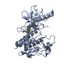
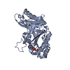
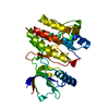
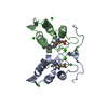
| Title | Crystal structure of 3'(2'),5'-bisphosphate nucleotidase1 from Entamoeba histolytica in complex with AMP and metal ions | ||||||
 Components Components | 3'(2'),5'-bisphosphate nucleotidase, putative | ||||||
 Keywords Keywords | HYDROLASE / Hydrolysis / adenosine 3' / 5'-bisphosphate (PAP) binding / Dephosphorylation | ||||||
| Function / homology |  Function and homology information Function and homology informationinositol-1,4-bisphosphate 1-phosphatase / inositol-1,4-bisphosphate 1-phosphatase activity / 3'(2'),5'-bisphosphate nucleotidase / 3'(2'),5'-bisphosphate nucleotidase activity / sulfate assimilation / phosphatidylinositol phosphate biosynthetic process / nucleotide binding / magnesium ion binding / cytoplasm Similarity search - Function | ||||||
| Biological species |   Entamoeba histolytica (eukaryote) Entamoeba histolytica (eukaryote) | ||||||
| Method |  X-RAY DIFFRACTION / MOLECULAR REPLACEMENT / Resolution: 2.6 Å X-RAY DIFFRACTION / MOLECULAR REPLACEMENT / Resolution: 2.6 Å | ||||||
 Authors Authors | Tarique, K.F. / Abdul Rehman, S.A. / Gourinath, S. | ||||||
 Citation Citation | Journal: Acta Crystallogr.,Sect.D / Year: 2014 Title: Structural elucidation of a dual-activity PAP phosphatase-1 from Entamoeba histolytica capable of hydrolysing both 3'-phosphoadenosine 5'-phosphate and inositol 1,4-bisphosphate Authors: Faisal Tarique, K. / Arif Abdul Rehman, S. / Gourinath, S. | ||||||
| History |
|
- Structure visualization
| Structure viewer | Molecule: MolmilJmol/JSmol |
|---|
- Downloads & links
-Download
| PDBx/mmCIF format | 4hxv.cif.gz | 75.8 KB | Display | PDBx/mmCIF format |
|---|---|---|---|---|
| PDB format | pdb4hxv.ent.gz | 54.7 KB | Display | PDB format |
| PDBx/mmJSON format | 4hxv.json.gz | Tree view | PDBx/mmJSON format | |
| Others |  Other downloads Other downloads |
-Validation report
| Arichive directory | https://data.pdbj.org/pub/pdb/validation_reports/hx/4hxvftp://data.pdbj.org/pub/pdb/validation_reports/hx/4hxv | HTTPS FTP |
|---|
-Related structure data
| Related structure data | 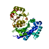 4o7iC  4gdg S: Starting model for refinement C: citing same article ( |
|---|---|
| Similar structure data |


-Links
 PDBj
PDBj


- Assembly
| Deposited unit | 
| ||||||||
|---|---|---|---|---|---|---|---|---|---|
| 1 |
| ||||||||
| Unit cell |
|
-Components
| #1: Protein | Mass: 36154.023 Da / Num. of mol.: 1 Source method: isolated from a genetically manipulated source Source: (gene. exp.) Entamoeba histolytica (eukaryote) / Strain: HM-1:IMSS / Gene: EHI_193350 / Plasmid: pET28(b) / Production host:  References: UniProt: C4M4T9, 3'(2'),5'-bisphosphate nucleotidase |
|---|---|
| #2: Chemical | ChemComp-MG /   Mass: 24.305 Da / Num. of mol.: 1 / Source method: obtained synthetically / Formula: Mg Mass: 24.305 Da / Num. of mol.: 1 / Source method: obtained synthetically / Formula: Mg |
| #3: Chemical | ChemComp-AMP /   Mass: 347.221 Da / Num. of mol.: 1 / Source method: obtained synthetically / Formula: C10H14N5O7P / Comment: AMP*YM Mass: 347.221 Da / Num. of mol.: 1 / Source method: obtained synthetically / Formula: C10H14N5O7P / Comment: AMP*YM |
| #4: Chemical | ChemComp-NA /   Mass: 22.990 Da / Num. of mol.: 1 / Source method: obtained synthetically / Formula: Na Mass: 22.990 Da / Num. of mol.: 1 / Source method: obtained synthetically / Formula: Na |
| #5: Water | ChemComp-HOH /  Mass: 18.015 Da / Num. of mol.: 47 / Source method: isolated from a natural source / Formula: H2O Mass: 18.015 Da / Num. of mol.: 47 / Source method: isolated from a natural source / Formula: H2O |
-Experimental details
-Experiment
| Experiment | Method: X-RAY DIFFRACTION / Number of used crystals: 1 |
|---|
- Sample preparation
| Crystal | Density Matthews: 2.06 Å3/Da / Density % sol: 40.35 % |
|---|---|
| Crystal grow | Temperature: 289 K / Method: vapor diffusion, hanging drop / pH: 7.5 Details: 28% PEG4000, 200mM NaCl, 5mM MgCl2, 5mM adenosine monophosphate (AMP), pH 7.5, VAPOR DIFFUSION, HANGING DROP, temperature 289K |
-Data collection
| Diffraction | Mean temperature: 100 K |
|---|---|
| Diffraction source | Source: ROTATING ANODE / Type: BRUKER AXS MICROSTAR / Wavelength: 1.54 Å |
| Detector | Type: MAR scanner 345 mm plate / Detector: IMAGE PLATE / Date: Aug 10, 2011 |
| Radiation | Protocol: SINGLE WAVELENGTH / Monochromatic (M) / Laue (L): M / Scattering type: x-ray |
| Radiation wavelength | Wavelength: 1.54 Å / Relative weight: 1 |
| Reflection | Resolution: 2.59→50 Å / Num. obs: 9190 / % possible obs: 98.9 % / Observed criterion σ(F): 0 / Observed criterion σ(I): -3 / Redundancy: 11.6 % / Rmerge(I) obs: 0.185 |
| Reflection shell | Resolution: 2.59→2.68 Å / Redundancy: 10.8 % / Rmerge(I) obs: 0.43 / Mean I/σ(I) obs: 5.319 / Num. unique all: 851 / % possible all: 88.8 |
- Processing
| Software |
| |||||||||||||||||||||||||||||||||||||||||||||
|---|---|---|---|---|---|---|---|---|---|---|---|---|---|---|---|---|---|---|---|---|---|---|---|---|---|---|---|---|---|---|---|---|---|---|---|---|---|---|---|---|---|---|---|---|---|---|
| Refinement | Method to determine structure: MOLECULAR REPLACEMENT Starting model: 4GDG 4gdg Resolution: 2.6→43.31 Å / Cor.coef. Fo:Fc: 0.903 / Cor.coef. Fo:Fc free: 0.849 / Occupancy max: 1 / Occupancy min: 0.5 / SU B: 13.459 / SU ML: 0.285 / Cross valid method: THROUGHOUT / σ(F): 0 / ESU R Free: 0.394 / Stereochemistry target values: MAXIMUM LIKELIHOOD / Details: HYDROGENS HAVE BEEN USED IF PRESENT IN THE INPUT
| |||||||||||||||||||||||||||||||||||||||||||||
| Solvent computation | Ion probe radii: 0.8 Å / Shrinkage radii: 0.8 Å / VDW probe radii: 1.2 Å / Solvent model: MASK | |||||||||||||||||||||||||||||||||||||||||||||
| Displacement parameters | Biso max: 40.39 Å2 / Biso mean: 22.2094 Å2 / Biso min: 8.48 Å2
| |||||||||||||||||||||||||||||||||||||||||||||
| Refine analyze | Luzzati coordinate error obs: 0.3455 Å | |||||||||||||||||||||||||||||||||||||||||||||
| Refinement step | Cycle: LAST / Resolution: 2.6→43.31 Å
| |||||||||||||||||||||||||||||||||||||||||||||
| Refine LS restraints |
| |||||||||||||||||||||||||||||||||||||||||||||
| LS refinement shell | Resolution: 2.602→2.67 Å / Total num. of bins used: 20
|
