- PDB-4hxc: Crystal structure of a putative glycosyl hydrolase (BACUNI_00951)... -
+
Open data
ID or keywords:
Loading...
-
Basic information
Entry
Database: PDB / ID: 4hxc
Title
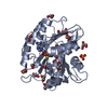
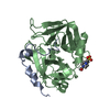
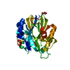
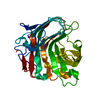
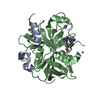
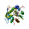
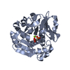
Crystal structure of a putative glycosyl hydrolase (BACUNI_00951) from Bacteroides uniformis ATCC 8492 at 2.15 A resolution
Components
putative glycosyl hydrolase
Keywords
HYDROLASE / PF06439 family protein / DUF1080 / Structural Genomics / Joint Center for Structural Genomics / JCSG / Protein Structure Initiative / PSI-BIOLOGY
Mass: 18.015 Da / Num. of mol.: 184 / Source method: isolated from a natural source / Formula: H2O
Has protein modification
Y
Sequence details
THE CONSTRUCT (RESIDUES 30-291) WAS EXPRESSED WITH A PURIFICATION TAG MGSDKIHHHHHHENLYFQG. THE TAG ...THE CONSTRUCT (RESIDUES 30-291) WAS EXPRESSED WITH A PURIFICATION TAG MGSDKIHHHHHHENLYFQG. THE TAG WAS REMOVED WITH TEV PROTEASE LEAVING ONLY A GLYCINE (0) FOLLOWED BY THE TARGET SEQUENCE.
-
Experimental details
-
Experiment
Experiment
Method: X-RAY DIFFRACTION / Number of used crystals: 1
-
Sample preparation
Crystal
Density Matthews: 3.53 Å3/Da / Density % sol: 65.13 %
Crystal grow
Temperature: 277 K / Method: vapor diffusion, sitting drop Details: 0.2M potassium fluoride 20% polyethylene glycol 3350, NANODROP, VAPOR DIFFUSION, SITTING DROP, temperature 277K
Type: DECTRIS PILATUS 6M / Detector: PIXEL / Date: Jun 28, 2012 Details: Flat mirror (vertical focusing); single crystal Si(111) bent monochromator (horizontal focusing)
Radiation
Monochromator: single crystal Si(111) bent / Protocol: MAD / Monochromatic (M) / Laue (L): M / Scattering type: x-ray
Radiation wavelength
ID
Wavelength (Å)
Relative weight
1
0.91837
1
2
0.9792
1
3
0.97868
1
Reflection
Resolution: 2.15→29.461 Å / Num. obs: 22690 / % possible obs: 93 % / Observed criterion σ(I): -3 / Biso Wilson estimate: 31.567 Å2 / Rmerge(I) obs: 0.073 / Net I/σ(I): 9.33
Reflection shell
Diffraction-ID: 1
Resolution (Å)
Highest resolution (Å)
Rmerge(I) obs
Mean I/σ(I) obs
Num. measured obs
Num. unique obs
% possible all
2.15-2.23
0.421
2.1
7635
4010
87.7
2.23-2.32
0.394
2.3
7594
4047
92.5
2.32-2.42
0.29
3
7424
3802
91.6
2.42-2.55
0.197
4.4
8662
4301
95.8
2.55-2.71
0.164
5.2
8301
4142
94.8
2.71-2.92
0.12
6.9
8322
4255
96.4
2.92-3.21
0.075
9.9
7715
3974
92
3.21-3.67
0.047
15.1
8369
4121
94.6
3.67-4.61
0.036
20.5
7593
3945
90.6
4.61
0.027
23.6
8611
4236
94.4
-
Phasing
Phasing
Method: MAD
-
Processing
Software
Name
Version
Classification
NB
MolProbity
3beta29
modelbuilding
PDB_EXTRACT
3.1
dataextraction
SHELX
phasing
SHARP
phasing
XSCALE
March15, 2012
datascaling
BUSTER-TNT
2.10.0
refinement
XDS
datareduction
SHELXD
phasing
BUSTER
2.10.0
refinement
Refinement
Method to determine structure: MAD / Resolution: 2.15→29.461 Å / Cor.coef. Fo:Fc: 0.9443 / Cor.coef. Fo:Fc free: 0.9201 / Occupancy max: 1 / Occupancy min: 0.5 / Cross valid method: THROUGHOUT / σ(F): 0 Details: 1. A MET-INHIBITION PROTOCOL WAS USED FOR SELENOMETHIONINE INCORPORATION DURING PROTEIN EXPRESSION. THE OCCUPANCY OF THE SE ATOMS IN THE MSE RESIDUES WAS REDUCED TO 0.75 FOR THE REDUCED ...Details: 1. A MET-INHIBITION PROTOCOL WAS USED FOR SELENOMETHIONINE INCORPORATION DURING PROTEIN EXPRESSION. THE OCCUPANCY OF THE SE ATOMS IN THE MSE RESIDUES WAS REDUCED TO 0.75 FOR THE REDUCED SCATTERING POWER DUE TO PARTIAL S-MET INCORPORATION. 2. ATOM RECORD CONTAINS SUM OF TLS AND RESIDUAL B FACTORS. ANISOU RECORD CONTAINS SUM OF TLS AND RESIDUAL U FACTORS. 3. THE MAD PHASES WERE USED AS RESTRAINTS DURING REFINEMENT. 4.POLYETHYLENE GLYCOL FRAGMENTS (PGE,PEG) FROM THE CRYSTALLIZATION AND 1,2-ETHANEDIOL (EDO),USED AS A CRYOPROTECTANT HAVE BEEN MODELED INTO THE STRUCTURE.
In the structure databanks used in Yorodumi, some data are registered as the other names, "COVID-19 virus" and "2019-nCoV". Here are the details of the virus and the list of structure data.
Jan 31, 2019. EMDB accession codes are about to change! (news from PDBe EMDB page)
EMDB accession codes are about to change! (news from PDBe EMDB page)
The allocation of 4 digits for EMDB accession codes will soon come to an end. Whilst these codes will remain in use, new EMDB accession codes will include an additional digit and will expand incrementally as the available range of codes is exhausted. The current 4-digit format prefixed with “EMD-” (i.e. EMD-XXXX) will advance to a 5-digit format (i.e. EMD-XXXXX), and so on. It is currently estimated that the 4-digit codes will be depleted around Spring 2019, at which point the 5-digit format will come into force.
The EM Navigator/Yorodumi systems omit the EMD- prefix.
Related info.:Q: What is EMD? / ID/Accession-code notation in Yorodumi/EM Navigator
Yorodumi is a browser for structure data from EMDB, PDB, SASBDB, etc.
This page is also the successor to EM Navigator detail page, and also detail information page/front-end page for Omokage search.
The word "yorodu" (or yorozu) is an old Japanese word meaning "ten thousand". "mi" (miru) is to see.
Related info.:EMDB / PDB / SASBDB / Comparison of 3 databanks / Yorodumi Search / Aug 31, 2016. New EM Navigator & Yorodumi / Yorodumi Papers / Jmol/JSmol / Function and homology information / Changes in new EM Navigator and Yorodumi
 Movie
Movie Controller

 Yorodumi
 Open data
 Basic information
 Components
 Keywords
 Function and homology information
 Bacteroides uniformis (bacteria)
 X-RAY DIFFRACTION /
  Authors
 Citation
 Structure visualization
 Downloads & links
 Other downloads


 PDBj
PDBj


 Assembly

 Mass: 62.068 Da / Num. of mol.: 3 / Source method: obtained synthetically / Formula: C2H6O2
 Mass: 150.173 Da / Num. of mol.: 1 / Source method: obtained synthetically / Formula: C6H14O4
 Mass: 106.120 Da / Num. of mol.: 1 / Source method: obtained synthetically / Formula: C4H10O3
 Mass: 18.015 Da / Num. of mol.: 184 / Source method: isolated from a natural source / Formula: H2O
 Sample preparation
 / Beamline: BL11-1 / Wavelength: 0.91837,0.9792,0.97868
 Processing