 Movie
Movie Controller

[English] 日本語
 Yorodumi
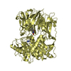
- PDB-4hvt: Structure of a Post-proline cleaving enzyme from Rickettsia typhi -
+ Open data

- Basic information
| Entry | Database: PDB / ID: 4hvt | ||||||
|---|---|---|---|---|---|---|---|
| Title | Structure of a Post-proline cleaving enzyme from Rickettsia typhi | ||||||
 Components Components | Post-proline cleaving enzyme | ||||||
 Keywords Keywords | HYDROLASE / SSGCID / Post-proline cleaving enzyme / Structural Genomics / Seattle Structural Genomics Center for Infectious Disease / post-proline cleavage protein | ||||||
| Function / homology |  Function and homology information Function and homology informationprolyl oligopeptidase / oligopeptidase activity / serine-type endopeptidase activity / proteolysis / cytosol Similarity search - Function | ||||||
| Biological species |  Rickettsia typhi (bacteria) Rickettsia typhi (bacteria) | ||||||
| Method |  X-RAY DIFFRACTION / SYNCHROTRON / SAD, MOLECULAR REPLACEMENT / SAD / molecular replacement / Resolution: 1.7 Å X-RAY DIFFRACTION / SYNCHROTRON / SAD, MOLECULAR REPLACEMENT / SAD / molecular replacement / Resolution: 1.7 Å | ||||||
 Authors Authors | Seattle Structural Genomics Center for Infectious Disease (SSGCID) | ||||||
 Citation Citation | Journal: To be Published Title: Structure of a Post-proline cleaving enzyme from Rickettsia typhi Authors: Seattle Structural Genomics Center for Infectious Disease (SSGCID) / Abendroth, J. / Sankaran, B. / Fox III, D. / Edwards, T.E. / Staker, B.L. | ||||||
| History |
|
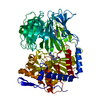
- Structure visualization
| Structure viewer | Molecule: MolmilJmol/JSmol |
|---|
- Downloads & links
-Download
| PDBx/mmCIF format | 4hvt.cif.gz | 303.1 KB | Display | PDBx/mmCIF format |
|---|---|---|---|---|
| PDB format | pdb4hvt.ent.gz | 244.1 KB | Display | PDB format |
| PDBx/mmJSON format | 4hvt.json.gz | Tree view | PDBx/mmJSON format | |
| Others |  Other downloads Other downloads |
-Validation report
| Arichive directory | https://data.pdbj.org/pub/pdb/validation_reports/hv/4hvtftp://data.pdbj.org/pub/pdb/validation_reports/hv/4hvt | HTTPS FTP |
|---|
-Related structure data
| Similar structure data | |
|---|---|
| Other databases |


-Links
 PDBj
PDBj
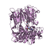
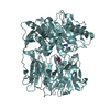
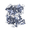
- Assembly
| Deposited unit | 
| ||||||||
|---|---|---|---|---|---|---|---|---|---|
| 1 |
| ||||||||
| Unit cell |
| ||||||||
| Components on special symmetry positions |
|
-Components
| #1: Protein | Mass: 82226.664 Da / Num. of mol.: 1 / Fragment: N-terminal truncation (UNP residues 19-720) Source method: isolated from a genetically manipulated source Source: (gene. exp.) Rickettsia typhi (bacteria) / Strain: ATCC VR-144 / Wilmington / Gene: ppcE, RT0165 / Plasmid: RityA.17583.b.B2 / Production host: | ||||
|---|---|---|---|---|---|
| #2: Chemical | ChemComp-EDO /   Mass: 62.068 Da / Num. of mol.: 6 / Source method: obtained synthetically / Formula: C2H6O2 Mass: 62.068 Da / Num. of mol.: 6 / Source method: obtained synthetically / Formula: C2H6O2#3: Chemical |   Mass: 35.453 Da / Num. of mol.: 2 / Source method: obtained synthetically / Formula: Cl Mass: 35.453 Da / Num. of mol.: 2 / Source method: obtained synthetically / Formula: Cl#4: Water | ChemComp-HOH / |  Mass: 18.015 Da / Num. of mol.: 746 / Source method: isolated from a natural source / Formula: H2O Mass: 18.015 Da / Num. of mol.: 746 / Source method: isolated from a natural source / Formula: H2O |
-Experimental details
-Experiment
| Experiment | Method: X-RAY DIFFRACTION / Number of used crystals: 2 |
|---|
- Sample preparation
| Crystal |
| |||||||||||||||
|---|---|---|---|---|---|---|---|---|---|---|---|---|---|---|---|---|
| Crystal grow |
|
-Data collection
| Diffraction |
| |||||||||||||||||||||||||||||||||||||||||||||||||||||||||||||||||||||||||||||||||||||||||||||||||||||||||||||||||||||||||||||||||||||||||||||||||||
|---|---|---|---|---|---|---|---|---|---|---|---|---|---|---|---|---|---|---|---|---|---|---|---|---|---|---|---|---|---|---|---|---|---|---|---|---|---|---|---|---|---|---|---|---|---|---|---|---|---|---|---|---|---|---|---|---|---|---|---|---|---|---|---|---|---|---|---|---|---|---|---|---|---|---|---|---|---|---|---|---|---|---|---|---|---|---|---|---|---|---|---|---|---|---|---|---|---|---|---|---|---|---|---|---|---|---|---|---|---|---|---|---|---|---|---|---|---|---|---|---|---|---|---|---|---|---|---|---|---|---|---|---|---|---|---|---|---|---|---|---|---|---|---|---|---|---|---|---|
| Diffraction source |
| |||||||||||||||||||||||||||||||||||||||||||||||||||||||||||||||||||||||||||||||||||||||||||||||||||||||||||||||||||||||||||||||||||||||||||||||||||
| Detector |
| |||||||||||||||||||||||||||||||||||||||||||||||||||||||||||||||||||||||||||||||||||||||||||||||||||||||||||||||||||||||||||||||||||||||||||||||||||
| Radiation |
| |||||||||||||||||||||||||||||||||||||||||||||||||||||||||||||||||||||||||||||||||||||||||||||||||||||||||||||||||||||||||||||||||||||||||||||||||||
| Radiation wavelength |
| |||||||||||||||||||||||||||||||||||||||||||||||||||||||||||||||||||||||||||||||||||||||||||||||||||||||||||||||||||||||||||||||||||||||||||||||||||
| Reflection | Number: 305759 / Rmerge(I) obs: 0.05 / D res high: 2.1 Å / Num. obs: 84077 / % possible obs: 99.3 | |||||||||||||||||||||||||||||||||||||||||||||||||||||||||||||||||||||||||||||||||||||||||||||||||||||||||||||||||||||||||||||||||||||||||||||||||||
| Diffraction reflection shell |
| |||||||||||||||||||||||||||||||||||||||||||||||||||||||||||||||||||||||||||||||||||||||||||||||||||||||||||||||||||||||||||||||||||||||||||||||||||
| Reflection | Resolution: 1.7→50 Å / Num. all: 87031 / Num. obs: 86126 / % possible obs: 99 % / Observed criterion σ(F): 0 / Observed criterion σ(I): -3 / Redundancy: 4.8 % / Biso Wilson estimate: 23.221 Å2 / Rmerge(I) obs: 0.065 / Net I/σ(I): 16.86 | |||||||||||||||||||||||||||||||||||||||||||||||||||||||||||||||||||||||||||||||||||||||||||||||||||||||||||||||||||||||||||||||||||||||||||||||||||
| Reflection shell |
|

-Phasing
| Phasing |
| |||||||||
|---|---|---|---|---|---|---|---|---|---|---|
| Phasing MAD | D res high: 2.1 Å / D res low: 46.03 Å / FOM : 0.416 / FOM acentric: 0.429 / FOM centric: 0.128 / Reflection: 43000 / Reflection acentric: 41043 / Reflection centric: 1776 | |||||||||
| Phasing MR | Model details: Phaser MODE: MR_AUTO
|
- Processing
| Software |
| ||||||||||||||||||||||||||||||||||||||||||||||||||||||||||||
|---|---|---|---|---|---|---|---|---|---|---|---|---|---|---|---|---|---|---|---|---|---|---|---|---|---|---|---|---|---|---|---|---|---|---|---|---|---|---|---|---|---|---|---|---|---|---|---|---|---|---|---|---|---|---|---|---|---|---|---|---|---|
| Refinement | Method to determine structure: SAD, MOLECULAR REPLACEMENT Starting model: An initial model was obtained from iodide SAD phasing using crystal 2 (P21). The initial model was then used as search model for C2 high resolution data set, crystal 1. Resolution: 1.7→50 Å / Cor.coef. Fo:Fc: 0.958 / Cor.coef. Fo:Fc free: 0.941 / WRfactor Rfree: 0.1996 / WRfactor Rwork: 0.1636 / Occupancy max: 1 / Occupancy min: 0.4 / FOM work R set: 0.8714 / SU B: 3.886 / SU ML: 0.067 / SU R Cruickshank DPI: 0.099 / SU Rfree: 0.1011 / Isotropic thermal model: isotropic, TLS / Cross valid method: THROUGHOUT / σ(F): 0 / ESU R: 0.099 / ESU R Free: 0.101 / Stereochemistry target values: MAXIMUM LIKELIHOOD Details: HYDROGENS HAVE BEEN ADDED IN THE RIDING POSITIONS. U VALUES: WITH TLS ADDED
| ||||||||||||||||||||||||||||||||||||||||||||||||||||||||||||
| Solvent computation | Ion probe radii: 0.8 Å / Shrinkage radii: 0.8 Å / VDW probe radii: 1.2 Å / Solvent model: MASK | ||||||||||||||||||||||||||||||||||||||||||||||||||||||||||||
| Displacement parameters | Biso max: 54.98 Å2 / Biso mean: 19.2341 Å2 / Biso min: 7.19 Å2
| ||||||||||||||||||||||||||||||||||||||||||||||||||||||||||||
| Refinement step | Cycle: LAST / Resolution: 1.7→50 Å
| ||||||||||||||||||||||||||||||||||||||||||||||||||||||||||||
| Refine LS restraints |
| ||||||||||||||||||||||||||||||||||||||||||||||||||||||||||||
| LS refinement shell | Resolution: 1.7→1.744 Å / Total num. of bins used: 20
| ||||||||||||||||||||||||||||||||||||||||||||||||||||||||||||
| Refinement TLS params. | Method: refined / Origin x: 23.725 Å / Origin y: 61.672 Å / Origin z: 16.053 Å
| ||||||||||||||||||||||||||||||||||||||||||||||||||||||||||||
| Refinement TLS group |
|
