 Movie
Movie Controller


+ Open data

- Basic information
| Entry | Database: PDB / ID: 4hts | ||||||
|---|---|---|---|---|---|---|---|
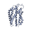
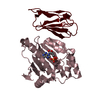
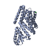
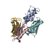
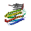
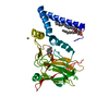
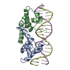
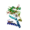
| Title | Crystal Structure of Twin Arginine Translocase Receptor- TatC | ||||||
 Components Components | Sec-independent protein translocase protein TatC | ||||||
 Keywords Keywords | PROTEIN TRANSPORT / Twin arginine translocase receptor | ||||||
| Function / homology | Sec-independent periplasmic protein translocase, conserved site / TatC family signature. / Sec-independent periplasmic protein translocase TatC / Sec-independent protein translocase protein (TatC) / proton motive force dependent protein transmembrane transporter activity / TAT protein transport complex / protein transport by the Tat complex / intracellular protein transmembrane transport / Sec-independent protein translocase protein TatC Function and homology information Function and homology information | ||||||
| Biological species |   Aquifex aeolicus VF5 (bacteria) Aquifex aeolicus VF5 (bacteria) | ||||||
| Method |  X-RAY DIFFRACTION / SYNCHROTRON / MOLECULAR REPLACEMENT / Resolution: 4 Å X-RAY DIFFRACTION / SYNCHROTRON / MOLECULAR REPLACEMENT / Resolution: 4 Å | ||||||
 Authors Authors | Ramasamy, S. / Chartron, J.W. / Clemons Jr., W.M. | ||||||
 Citation Citation | Journal: Structure / Year: 2013 Title: The Glove-like Structure of the Conserved Membrane Protein TatC Provides Insight into Signal Sequence Recognition in Twin-Arginine Translocation. Authors: Ramasamy, S. / Abrol, R. / Suloway, C.J. / Clemons, W.M. | ||||||
| History |
|
- Structure visualization
| Structure viewer | Molecule: MolmilJmol/JSmol |
|---|
- Downloads & links
-Download
| PDBx/mmCIF format | 4hts.cif.gz | 51.5 KB | Display | PDBx/mmCIF format |
|---|---|---|---|---|
| PDB format | pdb4hts.ent.gz | 38.4 KB | Display | PDB format |
| PDBx/mmJSON format | 4hts.json.gz | Tree view | PDBx/mmJSON format | |
| Others |  Other downloads Other downloads |
-Validation report
| Arichive directory | https://data.pdbj.org/pub/pdb/validation_reports/ht/4htsftp://data.pdbj.org/pub/pdb/validation_reports/ht/4hts | HTTPS FTP |
|---|
-Related structure data


-Links
 PDBj
PDBj- Assembly
| Deposited unit | 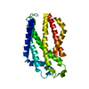
| ||||||||
|---|---|---|---|---|---|---|---|---|---|
| 1 |
| ||||||||
| Unit cell |
|
-Components
| #1: Protein | Mass: 26436.027 Da / Num. of mol.: 1 / Fragment: UNP residues 1-232 / Mutation: K40A, E41A Source method: isolated from a genetically manipulated source Source: (gene. exp.) Aquifex aeolicus VF5 (bacteria) / Strain: VF5 / Gene: tatC, aq_1267 / Plasmid: pET33b / Production host: |
|---|
-Experimental details
-Experiment
| Experiment | Method: X-RAY DIFFRACTION / Number of used crystals: 1 |
|---|
- Sample preparation
| Crystal | Density Matthews: 6.2 Å3/Da / Density % sol: 80.15 % |
|---|---|
| Crystal grow | Temperature: 293 K / Method: vapor diffusion, sitting drop / pH: 7.5 Details: 28% Jeffamine ED-2001 0.05M HEPES pH 7.5 10% Methyl-2,4-pentanediol (MPD)., VAPOR DIFFUSION, SITTING DROP, temperature 293K |
-Data collection
| Diffraction | Mean temperature: 100 K | |||||||||||||||||||||||||||||||||||||||||||||||||||||||
|---|---|---|---|---|---|---|---|---|---|---|---|---|---|---|---|---|---|---|---|---|---|---|---|---|---|---|---|---|---|---|---|---|---|---|---|---|---|---|---|---|---|---|---|---|---|---|---|---|---|---|---|---|---|---|---|---|
| Diffraction source | Source: SYNCHROTRON / Site: SSRL  / Beamline: BL12-2 / Wavelength: 1.08 Å / Beamline: BL12-2 / Wavelength: 1.08 Å | |||||||||||||||||||||||||||||||||||||||||||||||||||||||
| Detector | Type: MARMOSAIC 325 mm CCD / Detector: CCD / Date: Nov 25, 2008 | |||||||||||||||||||||||||||||||||||||||||||||||||||||||
| Radiation | Monochromator: LIQUID NITROGEN-COOLED DOUBLE CRYSTAL / Protocol: SINGLE WAVELENGTH / Monochromatic (M) / Laue (L): M / Scattering type: x-ray | |||||||||||||||||||||||||||||||||||||||||||||||||||||||
| Radiation wavelength | Wavelength: 1.08 Å / Relative weight: 1 | |||||||||||||||||||||||||||||||||||||||||||||||||||||||
| Reflection | Resolution: 4→110.43 Å / Num. obs: 5887 / % possible obs: 99.77 % / Observed criterion σ(F): 22.03 / Observed criterion σ(I): 66.49 / Redundancy: 7.7 % / Rsym value: 0.062 / Net I/σ(I): 16.8 | |||||||||||||||||||||||||||||||||||||||||||||||||||||||
| Reflection shell |
|
- Processing
| Software |
| |||||||||||||||||||||||||||||||||||
|---|---|---|---|---|---|---|---|---|---|---|---|---|---|---|---|---|---|---|---|---|---|---|---|---|---|---|---|---|---|---|---|---|---|---|---|---|
| Refinement | Method to determine structure: MOLECULAR REPLACEMENT / Resolution: 4→29.911 Å / Cor.coef. Fo:Fc: 0.867 / Cor.coef. Fo:Fc free: 0.829 / SU ML: 0.54 / σ(F): 1.91 / Phase error: 39.28 / Stereochemistry target values: ML Details: HYDROGENS HAVE BEEN ADDED IN THE RIDING POSITIONS U VALUES : REFINED INDIVIDUALLY
| |||||||||||||||||||||||||||||||||||
| Solvent computation | Shrinkage radii: 0.9 Å / VDW probe radii: 1.11 Å / Solvent model: FLAT BULK SOLVENT MODEL | |||||||||||||||||||||||||||||||||||
| Displacement parameters | Biso mean: 211.998 Å2
| |||||||||||||||||||||||||||||||||||
| Refinement step | Cycle: LAST / Resolution: 4→29.911 Å
| |||||||||||||||||||||||||||||||||||
| Refine LS restraints |
| |||||||||||||||||||||||||||||||||||
| LS refinement shell |
|
