 Movie
Movie Controller


+ Open data

- Basic information
| Entry | Database: PDB / ID: 4hsu | ||||||
|---|---|---|---|---|---|---|---|
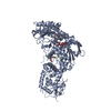
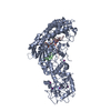
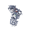
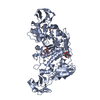
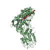
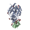
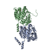
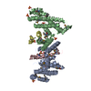
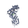
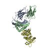
| Title | Crystal structure of LSD2-NPAC with H3(1-26)in space group P21 | ||||||
 Components Components |
| ||||||
 Keywords Keywords | OXIDOREDUCTASE / histone demethylase | ||||||
| Function / homology |  Function and homology information Function and homology informationepigenetic programing of female pronucleus / [histone H3]-N6,N6-dimethyl-L-lysine4 FAD-dependent demethylase / FAD-dependent H3K4me/H3K4me3 demethylase activity / genomic imprinting / transcription elongation-coupled chromatin remodeling / chromatin-protein adaptor activity / histone demethylase activity / nucleosome binding / NR1H3 & NR1H2 regulate gene expression linked to cholesterol transport and efflux / FAD binding ...epigenetic programing of female pronucleus / [histone H3]-N6,N6-dimethyl-L-lysine4 FAD-dependent demethylase / FAD-dependent H3K4me/H3K4me3 demethylase activity / genomic imprinting / transcription elongation-coupled chromatin remodeling / chromatin-protein adaptor activity / histone demethylase activity / nucleosome binding / NR1H3 & NR1H2 regulate gene expression linked to cholesterol transport and efflux / FAD binding / transcription initiation-coupled chromatin remodeling / HDMs demethylate histones / NAD binding / NADP binding / structural constituent of chromatin / UCH proteinases / nucleosome / histone binding / oxidoreductase activity / protein heterodimerization activity / chromatin binding / chromatin / DNA binding / zinc ion binding / nucleoplasm / nucleus / cytosol Similarity search - Function | ||||||
| Biological species |  Homo sapiens (human) Homo sapiens (human) | ||||||
| Method |  X-RAY DIFFRACTION / SYNCHROTRON / MOLECULAR REPLACEMENT / Resolution: 1.988 Å X-RAY DIFFRACTION / SYNCHROTRON / MOLECULAR REPLACEMENT / Resolution: 1.988 Å | ||||||
 Authors Authors | Chen, F. / Dong, Z. / Fang, J. / Xu, Y. | ||||||
 Citation Citation | Journal: Cell Res. / Year: 2013 Title: Structural insight into substrate recognition by histone demethylase LSD2/KDM1b. Authors: Chen, F. / Yang, H. / Dong, Z. / Fang, J. / Wang, P. / Zhu, T. / Gong, W. / Fang, R. / Shi, Y.G. / Li, Z. / Xu, Y. | ||||||
| History |
|
- Structure visualization
| Structure viewer | Molecule: MolmilJmol/JSmol |
|---|
- Downloads & links
-Download
| PDBx/mmCIF format | 4hsu.cif.gz | 181.6 KB | Display | PDBx/mmCIF format |
|---|---|---|---|---|
| PDB format | pdb4hsu.ent.gz | 136.6 KB | Display | PDB format |
| PDBx/mmJSON format | 4hsu.json.gz | Tree view | PDBx/mmJSON format | |
| Others |  Other downloads Other downloads |
-Validation report
| Arichive directory | https://data.pdbj.org/pub/pdb/validation_reports/hs/4hsuftp://data.pdbj.org/pub/pdb/validation_reports/hs/4hsu | HTTPS FTP |
|---|
-Related structure data
| Related structure data | 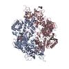 4gu0C 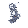 4gu1S S: Starting model for refinement C: citing same article ( |
|---|---|
| Similar structure data |
-Links
 PDBj
PDBj


- Assembly
| Deposited unit | 
| ||||||||
|---|---|---|---|---|---|---|---|---|---|
| 1 |
| ||||||||
| Unit cell |
|
-Components
-Protein , 2 types, 2 molecules AB
| #1: Protein | Mass: 87051.547 Da / Num. of mol.: 1 / Fragment: UNP residues 51-822 Source method: isolated from a genetically manipulated source Source: (gene. exp.) Homo sapiens (human) / Gene: LSD2 / Plasmid: modified pFastBac1 / Cell line (production host): sf9 / Production host:   Spodoptera frugiperda (fall armyworm) / References: UniProt: Q8NB78, Oxidoreductases Spodoptera frugiperda (fall armyworm) / References: UniProt: Q8NB78, Oxidoreductases |
|---|---|
| #2: Protein | Mass: 13528.271 Da / Num. of mol.: 1 / Fragment: UNP residues 152-268 Source method: isolated from a genetically manipulated source Source: (gene. exp.) Homo sapiens (human) / Gene: GLYR1 / Plasmid: pGEX-6P-1 / Production host:  |
-Protein/peptide , 1 types, 1 molecules C
| #3: Protein/peptide | Mass: 3177.750 Da / Num. of mol.: 1 / Fragment: UNP residues 2-31 / Mutation: K4M / Source method: obtained synthetically / Details: synthetic peptide / Source: (synth.) |
|---|
-Non-polymers , 3 types, 318 molecules 


| #4: Chemical | ChemComp-FAD /  Mass: 785.550 Da / Num. of mol.: 1 / Source method: obtained synthetically / Formula: C27H33N9O15P2 / Comment: FAD*YM Mass: 785.550 Da / Num. of mol.: 1 / Source method: obtained synthetically / Formula: C27H33N9O15P2 / Comment: FAD*YM | ||
|---|---|---|---|
| #5: Chemical |  Mass: 65.409 Da / Num. of mol.: 3 / Source method: obtained synthetically / Formula: Zn Mass: 65.409 Da / Num. of mol.: 3 / Source method: obtained synthetically / Formula: Zn#6: Water | ChemComp-HOH / | Mass: 18.015 Da / Num. of mol.: 314 / Source method: isolated from a natural source / Formula: H2O |
-Experimental details
-Experiment
| Experiment | Method: X-RAY DIFFRACTION / Number of used crystals: 1 |
|---|
- Sample preparation
| Crystal | Density Matthews: 2.33 Å3/Da / Density % sol: 47.22 % |
|---|---|
| Crystal grow | Temperature: 277 K / Method: vapor diffusion, hanging drop / pH: 6 Details: 0.02M Citric acid, 0.03M Bis_tris propane, 10% PEG3350, pH 6.0, VAPOR DIFFUSION, HANGING DROP, temperature 277K |
-Data collection
| Diffraction | Mean temperature: 100 K |
|---|---|
| Diffraction source | Source: SYNCHROTRON / Site: SSRF  / Beamline: BL17U / Wavelength: 0.9793 Å / Beamline: BL17U / Wavelength: 0.9793 Å |
| Detector | Type: ADSC QUANTUM 315r / Detector: CCD / Date: Apr 23, 2011 |
| Radiation | Monochromator: SAGITALLY FOCUSED Si(111) / Protocol: SINGLE WAVELENGTH / Monochromatic (M) / Laue (L): M / Scattering type: x-ray |
| Radiation wavelength | Wavelength: 0.9793 Å / Relative weight: 1 |
| Reflection | Resolution: 1.988→50 Å / Num. obs: 64821 / % possible obs: 98.99 % / Biso Wilson estimate: 29.89 Å2 |
- Processing
| Software |
| ||||||||||||||||||||||||||||||||||||||||||||||||||||||||||||||||||||||||||||||||||||||||||||||||||||||||||||||||||||||||||||||||||||||||||||||||||||||||||||||||||||||||
|---|---|---|---|---|---|---|---|---|---|---|---|---|---|---|---|---|---|---|---|---|---|---|---|---|---|---|---|---|---|---|---|---|---|---|---|---|---|---|---|---|---|---|---|---|---|---|---|---|---|---|---|---|---|---|---|---|---|---|---|---|---|---|---|---|---|---|---|---|---|---|---|---|---|---|---|---|---|---|---|---|---|---|---|---|---|---|---|---|---|---|---|---|---|---|---|---|---|---|---|---|---|---|---|---|---|---|---|---|---|---|---|---|---|---|---|---|---|---|---|---|---|---|---|---|---|---|---|---|---|---|---|---|---|---|---|---|---|---|---|---|---|---|---|---|---|---|---|---|---|---|---|---|---|---|---|---|---|---|---|---|---|---|---|---|---|---|---|---|---|
| Refinement | Method to determine structure: MOLECULAR REPLACEMENT Starting model: 4GU1 Resolution: 1.988→40.436 Å / Occupancy max: 1 / Occupancy min: 1 / FOM work R set: 0.7926 / SU ML: 0.23 / σ(F): 1.34 / Phase error: 27.39 / Stereochemistry target values: ML
| ||||||||||||||||||||||||||||||||||||||||||||||||||||||||||||||||||||||||||||||||||||||||||||||||||||||||||||||||||||||||||||||||||||||||||||||||||||||||||||||||||||||||
| Solvent computation | Shrinkage radii: 0.9 Å / VDW probe radii: 1.11 Å / Solvent model: FLAT BULK SOLVENT MODEL | ||||||||||||||||||||||||||||||||||||||||||||||||||||||||||||||||||||||||||||||||||||||||||||||||||||||||||||||||||||||||||||||||||||||||||||||||||||||||||||||||||||||||
| Displacement parameters | Biso max: 88.59 Å2 / Biso mean: 38.2711 Å2 / Biso min: 19.39 Å2 | ||||||||||||||||||||||||||||||||||||||||||||||||||||||||||||||||||||||||||||||||||||||||||||||||||||||||||||||||||||||||||||||||||||||||||||||||||||||||||||||||||||||||
| Refinement step | Cycle: LAST / Resolution: 1.988→40.436 Å
| ||||||||||||||||||||||||||||||||||||||||||||||||||||||||||||||||||||||||||||||||||||||||||||||||||||||||||||||||||||||||||||||||||||||||||||||||||||||||||||||||||||||||
| Refine LS restraints |
| ||||||||||||||||||||||||||||||||||||||||||||||||||||||||||||||||||||||||||||||||||||||||||||||||||||||||||||||||||||||||||||||||||||||||||||||||||||||||||||||||||||||||
| LS refinement shell | Refine-ID: X-RAY DIFFRACTION / Total num. of bins used: 23
|
