cellular response to heat / protein refolding / ATP hydrolysis activity / ATP binding / identical protein binding / cytoplasm Similarity search - Function
Chaperonin ClpB / ClpA/B, conserved site 2 / Chaperonins clpA/B signature 2. / ClpA/B, conserved site 1 / Chaperonins clpA/B signature 1. / ClpA/ClpB, AAA lid domain / AAA lid domain / : / Clp repeat (R) N-terminal domain / Clp repeat (R) domain profile. ...Chaperonin ClpB / ClpA/B, conserved site 2 / Chaperonins clpA/B signature 2. / ClpA/B, conserved site 1 / Chaperonins clpA/B signature 1. / ClpA/ClpB, AAA lid domain / AAA lid domain / : / Clp repeat (R) N-terminal domain / Clp repeat (R) domain profile. / Clp, repeat (R) domain / Clp, N-terminal domain superfamily / ClpA/B family / Clp ATPase, C-terminal / C-terminal, D2-small domain, of ClpB protein / C-terminal, D2-small domain, of ClpB protein / AAA domain (Cdc48 subfamily) / ATPase family associated with various cellular activities (AAA) / ATPase, AAA-type, core / P-loop containing nucleotide triphosphate hydrolases / ATPases associated with a variety of cellular activities / AAA+ ATPase domain / Rossmann fold / P-loop containing nucleoside triphosphate hydrolase / 3-Layer(aba) Sandwich / Alpha Beta Similarity search - Domain/homology
Resolution: 2.2→47.36 Å / Cor.coef. Fo:Fc: 0.939 / Cor.coef. Fo:Fc free: 0.918 / SU B: 13.583 / SU ML: 0.166 / Cross valid method: THROUGHOUT / ESU R: 0.272 / ESU R Free: 0.218 / Stereochemistry target values: MAXIMUM LIKELIHOOD / Details: HYDROGENS HAVE BEEN ADDED IN THE RIDING POSITIONS
Rfactor
Num. reflection
% reflection
Selection details
Rfree
0.26631
1207
5 %
RANDOM
Rwork
0.22536
-
-
-
obs
0.22742
23027
100 %
-
all
-
23027
-
-
Solvent computation
Ion probe radii: 0.8 Å / Shrinkage radii: 0.8 Å / VDW probe radii: 1.4 Å / Solvent model: MASK
Displacement parameters
Biso mean: 43.543 Å2
Baniso -1
Baniso -2
Baniso -3
1-
0.79 Å2
0.39 Å2
0 Å2
2-
-
0.79 Å2
0 Å2
3-
-
-
-1.18 Å2
Refinement step
Cycle: LAST / Resolution: 2.2→47.36 Å
Protein
Nucleic acid
Ligand
Solvent
Total
Num. atoms
2946
0
32
86
3064
Refine LS restraints
Refine-ID
Type
Dev ideal
Dev ideal target
Number
X-RAY DIFFRACTION
r_bond_refined_d
0.009
0.022
3041
X-RAY DIFFRACTION
r_angle_refined_deg
1.189
2.01
4101
X-RAY DIFFRACTION
r_dihedral_angle_1_deg
4.79
5
376
X-RAY DIFFRACTION
r_dihedral_angle_2_deg
34.102
23.529
153
X-RAY DIFFRACTION
r_dihedral_angle_3_deg
16.011
15
599
X-RAY DIFFRACTION
r_dihedral_angle_4_deg
16.866
15
40
X-RAY DIFFRACTION
r_chiral_restr
0.078
0.2
470
X-RAY DIFFRACTION
r_gen_planes_refined
0.004
0.021
2257
X-RAY DIFFRACTION
r_mcbond_it
0.459
1.5
1849
X-RAY DIFFRACTION
r_mcangle_it
0.923
2
2969
X-RAY DIFFRACTION
r_scbond_it
1.66
3
1192
X-RAY DIFFRACTION
r_scangle_it
2.95
4.5
1127
LS refinement shell
Resolution: 2.2→2.257 Å / Total num. of bins used: 20
Rfactor
Num. reflection
% reflection
Rfree
0.313
87
-
Rwork
0.258
1652
-
obs
-
1652
100 %
Refinement TLS params.
Method: refined / Refine-ID: X-RAY DIFFRACTION
ID
L11 (°2)
L12 (°2)
L13 (°2)
L22 (°2)
L23 (°2)
L33 (°2)
S11 (Å °)
S12 (Å °)
S13 (Å °)
S21 (Å °)
S22 (Å °)
S23 (Å °)
S31 (Å °)
S32 (Å °)
S33 (Å °)
T11 (Å2)
T12 (Å2)
T13 (Å2)
T22 (Å2)
T23 (Å2)
T33 (Å2)
Origin x (Å)
Origin y (Å)
Origin z (Å)
1
2.3009
-1.2189
-0.5502
1.7856
0.6543
1.479
-0.1019
-0.0232
-0.202
0.2353
0.0445
0.2932
-0.1185
-0.1845
0.0574
0.0713
0.0265
0.0433
0.2122
0.0535
0.0584
0.3563
16.5053
-14.8097
2
2.5917
-0.9135
0.4208
2.1456
-0.9272
1.243
-0.0944
-0.0216
-0.3089
0.19
-0.0097
-0.3236
0.0425
0.1785
0.1041
0.0629
-0.0179
-0.0313
0.3531
0.048
0.2048
35.3975
8.057
-24.5659
Refinement TLS group
ID
Refine-ID
Refine TLS-ID
Auth asym-ID
Auth seq-ID
1
X-RAY DIFFRACTION
1
A
151 - 395
2
X-RAY DIFFRACTION
2
A
396 - 532
+
About Yorodumi
-
News
-
Feb 9, 2022. New format data for meta-information of EMDB entries
New format data for meta-information of EMDB entries
Version 3 of the EMDB header file is now the official format.
The previous official version 1.9 will be removed from the archive.
In the structure databanks used in Yorodumi, some data are registered as the other names, "COVID-19 virus" and "2019-nCoV". Here are the details of the virus and the list of structure data.
Jan 31, 2019. EMDB accession codes are about to change! (news from PDBe EMDB page)
EMDB accession codes are about to change! (news from PDBe EMDB page)
The allocation of 4 digits for EMDB accession codes will soon come to an end. Whilst these codes will remain in use, new EMDB accession codes will include an additional digit and will expand incrementally as the available range of codes is exhausted. The current 4-digit format prefixed with “EMD-” (i.e. EMD-XXXX) will advance to a 5-digit format (i.e. EMD-XXXXX), and so on. It is currently estimated that the 4-digit codes will be depleted around Spring 2019, at which point the 5-digit format will come into force.
The EM Navigator/Yorodumi systems omit the EMD- prefix.
Related info.:Q: What is EMD? / ID/Accession-code notation in Yorodumi/EM Navigator
Yorodumi is a browser for structure data from EMDB, PDB, SASBDB, etc.
This page is also the successor to EM Navigator detail page, and also detail information page/front-end page for Omokage search.
The word "yorodu" (or yorozu) is an old Japanese word meaning "ten thousand". "mi" (miru) is to see.
Related info.:EMDB / PDB / SASBDB / Comparison of 3 databanks / Yorodumi Search / Aug 31, 2016. New EM Navigator & Yorodumi / Yorodumi Papers / Jmol/JSmol / Function and homology information / Changes in new EM Navigator and Yorodumi
 Movie
Movie Controller

 Yorodumi
 Open data
 Basic information
 Components
 Keywords
 Function and homology information
 Thermus thermophilus (bacteria)
 X-RAY DIFFRACTION /
  Authors
 Citation
 Structure visualization
 Downloads & links
 Other downloads


 PDBj
PDBj


 Assembly
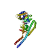
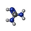
 Mass: 59.070 Da / Num. of mol.: 1 / Source method: obtained synthetically / Formula: CH5N3
 Mass: 427.201 Da / Num. of mol.: 1 / Source method: obtained synthetically / Formula: C10H15N5O10P2 / Comment: ADP, energy-carrying molecule*YM
 Mass: 35.453 Da / Num. of mol.: 1 / Source method: obtained synthetically / Formula: Cl
 Mass: 18.015 Da / Num. of mol.: 86 / Source method: isolated from a natural source / Formula: H2O
 Sample preparation
 / Beamline: X10SA / Wavelength: 0.9785 Å
 Processing