 Movie
Movie Controller

[English] 日本語
 Yorodumi
- PDB-4hs5: Frataxin from Psychromonas ingrahamii as a model to study stabili... -
+ Open data

- Basic information
| Entry | Database: PDB / ID: 4hs5 | ||||||
|---|---|---|---|---|---|---|---|
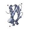
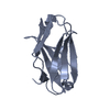
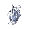
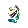
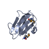
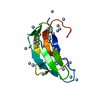
| Title | Frataxin from Psychromonas ingrahamii as a model to study stability modulation within CyaY protein family | ||||||
 Components Components | Protein CyaY | ||||||
 Keywords Keywords | METAL BINDING PROTEIN / pFXN / frataxin / CyaY / Psychromonas ingrahamii / Frataxin-like domain / CyaY protein family (pfam ID PF01491) / Iron homeostasis-redox balance | ||||||
| Function / homology |  Function and homology information Function and homology informationiron-sulfur cluster assembly / ferric iron binding / ferrous iron binding / cytosol Similarity search - Function | ||||||
| Biological species |  Psychromonas ingrahamii (bacteria) Psychromonas ingrahamii (bacteria) | ||||||
| Method |  X-RAY DIFFRACTION / SYNCHROTRON / MOLECULAR REPLACEMENT / Resolution: 1.45 Å X-RAY DIFFRACTION / SYNCHROTRON / MOLECULAR REPLACEMENT / Resolution: 1.45 Å | ||||||
 Authors Authors | Roman, E.A. / Cousido-siah, A. / Mitschler, A. / Podjarny, A. / Santos, J. | ||||||
 Citation Citation | Journal: Biochim.Biophys.Acta / Year: 2013 Title: Frataxin from Psychromonas ingrahamii as a model to study stability modulation within the CyaY protein family Authors: Roman, E.A. / Faraj, S.E. / Cousido-Siah, A. / Mitschler, A. / Podjarny, A. / Santos, J. | ||||||
| History |
|
- Structure visualization
| Structure viewer | Molecule: MolmilJmol/JSmol |
|---|
- Downloads & links
-Download
| PDBx/mmCIF format | 4hs5.cif.gz | 61.3 KB | Display | PDBx/mmCIF format |
|---|---|---|---|---|
| PDB format | pdb4hs5.ent.gz | 45 KB | Display | PDB format |
| PDBx/mmJSON format | 4hs5.json.gz | Tree view | PDBx/mmJSON format | |
| Others |  Other downloads Other downloads |
-Validation report
| Arichive directory | https://data.pdbj.org/pub/pdb/validation_reports/hs/4hs5ftp://data.pdbj.org/pub/pdb/validation_reports/hs/4hs5 | HTTPS FTP |
|---|
-Related structure data
| Related structure data |  1ew4S S: Starting model for refinement |
|---|---|
| Similar structure data |
-Links
 PDBj
PDBj
- Assembly
| Deposited unit | 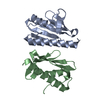
| ||||||||
|---|---|---|---|---|---|---|---|---|---|
| 1 | 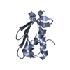
| ||||||||
| 2 | 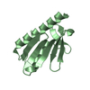
| ||||||||
| Unit cell |
|
-Components
| #1: Protein | Mass: 12295.387 Da / Num. of mol.: 2 Source method: isolated from a genetically manipulated source Details: Kanamycin resistance, IPTG inducible / Source: (gene. exp.) Psychromonas ingrahamii (bacteria) / Strain: 37 / Gene: cyaY, Ping_0042 / Plasmid: pJexpress411:56977 / Production host: #2: Water | ChemComp-HOH / |  Mass: 18.015 Da / Num. of mol.: 275 / Source method: isolated from a natural source / Formula: H2O Mass: 18.015 Da / Num. of mol.: 275 / Source method: isolated from a natural source / Formula: H2O |
|---|
-Experimental details
-Experiment
| Experiment | Method: X-RAY DIFFRACTION / Number of used crystals: 1 |
|---|
- Sample preparation
| Crystal | Density Matthews: 1.86 Å3/Da / Density % sol: 33.78 % |
|---|---|
| Crystal grow | Temperature: 297 K / Method: vapor diffusion, hanging drop / pH: 4.8 Details: 20mM sodium acetate, 200mM MgCl2, 29.5% PEG 4000, pH 4.80, VAPOR DIFFUSION, HANGING DROP, temperature 297K |
-Data collection
| Diffraction | Mean temperature: 177 K |
|---|---|
| Diffraction source | Source: SYNCHROTRON / Site: SLS  / Beamline: X06DA / Wavelength: 0.9191 Å / Beamline: X06DA / Wavelength: 0.9191 Å |
| Detector | Type: DECTRIS PILATUS 2M / Detector: PIXEL / Date: Apr 2, 2012 |
| Radiation | Monochromator: Bartels Monochromator / Protocol: SINGLE WAVELENGTH / Monochromatic (M) / Laue (L): M / Scattering type: x-ray |
| Radiation wavelength | Wavelength: 0.9191 Å / Relative weight: 1 |
| Reflection | Resolution: 1.45→50 Å / Num. all: 31292 / Num. obs: 31292 / % possible obs: 97.1 % / Observed criterion σ(F): 0 / Observed criterion σ(I): 0 / Redundancy: 3.4 % / Rmerge(I) obs: 0.036 / Net I/σ(I): 26.5 |
| Reflection shell | Resolution: 1.45→1.5 Å / % possible all: 97.1 |
- Processing
| Software | Name: PHENIX / Version: (phenix.refine: 1.8_1069) / Classification: refinement | ||||||||||||||||||||||||||||||||||||||||||||||||||||||||||||||||||||||||||||||||||||
|---|---|---|---|---|---|---|---|---|---|---|---|---|---|---|---|---|---|---|---|---|---|---|---|---|---|---|---|---|---|---|---|---|---|---|---|---|---|---|---|---|---|---|---|---|---|---|---|---|---|---|---|---|---|---|---|---|---|---|---|---|---|---|---|---|---|---|---|---|---|---|---|---|---|---|---|---|---|---|---|---|---|---|---|---|---|
| Refinement | Method to determine structure: MOLECULAR REPLACEMENT Starting model: PDB ID 1EW4 Resolution: 1.45→31.186 Å / Occupancy max: 1 / Occupancy min: 0 / FOM work R set: 0.8479 / SU ML: 0.17 / σ(F): 0 / Phase error: 22.53 / Stereochemistry target values: Engh & Huber
| ||||||||||||||||||||||||||||||||||||||||||||||||||||||||||||||||||||||||||||||||||||
| Solvent computation | Shrinkage radii: 0.9 Å / VDW probe radii: 1.11 Å / Solvent model: FLAT BULK SOLVENT MODEL | ||||||||||||||||||||||||||||||||||||||||||||||||||||||||||||||||||||||||||||||||||||
| Displacement parameters | Biso max: 58.38 Å2 / Biso mean: 15.7965 Å2 / Biso min: 2.92 Å2 | ||||||||||||||||||||||||||||||||||||||||||||||||||||||||||||||||||||||||||||||||||||
| Refinement step | Cycle: LAST / Resolution: 1.45→31.186 Å
| ||||||||||||||||||||||||||||||||||||||||||||||||||||||||||||||||||||||||||||||||||||
| Refine LS restraints |
| ||||||||||||||||||||||||||||||||||||||||||||||||||||||||||||||||||||||||||||||||||||
| LS refinement shell | Refine-ID: X-RAY DIFFRACTION / Total num. of bins used: 11
|
