 Movie
Movie Controller


+ Open data

- Basic information
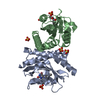
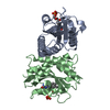
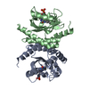
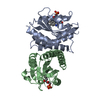
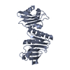
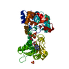
| Entry | Database: PDB / ID: 4hnb | ||||||
|---|---|---|---|---|---|---|---|
| Title | Crystal Structure of Rhodobacter Sphaeroides LOV protein | ||||||
 Components Components | LOV protein | ||||||
 Keywords Keywords | SIGNALING PROTEIN / LOV PAS HTH / signaling | ||||||
| Function / homology |  Function and homology information Function and homology information | ||||||
| Biological species |  Rhodobacter sphaeroides (bacteria) Rhodobacter sphaeroides (bacteria) | ||||||
| Method |  X-RAY DIFFRACTION / SYNCHROTRON / MOLECULAR REPLACEMENT / Resolution: 2.34 Å X-RAY DIFFRACTION / SYNCHROTRON / MOLECULAR REPLACEMENT / Resolution: 2.34 Å | ||||||
 Authors Authors | Crane, B.R. / Conrad, K.S. / Bilwes, A.M. | ||||||
 Citation Citation | Journal: Biochemistry / Year: 2013 Title: Light-induced subunit dissociation by a light-oxygen-voltage domain photoreceptor from Rhodobacter sphaeroides. Authors: Conrad, K.S. / Bilwes, A.M. / Crane, B.R. | ||||||
| History |
|
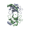
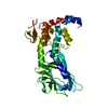
- Structure visualization
| Structure viewer | Molecule: MolmilJmol/JSmol |
|---|
- Downloads & links
-Download
| PDBx/mmCIF format | 4hnb.cif.gz | 83.4 KB | Display | PDBx/mmCIF format |
|---|---|---|---|---|
| PDB format | pdb4hnb.ent.gz | 63.8 KB | Display | PDB format |
| PDBx/mmJSON format | 4hnb.json.gz | Tree view | PDBx/mmJSON format | |
| Others |  Other downloads Other downloads |
-Validation report
| Arichive directory | https://data.pdbj.org/pub/pdb/validation_reports/hn/4hnbftp://data.pdbj.org/pub/pdb/validation_reports/hn/4hnb | HTTPS FTP |
|---|
-Related structure data


-Links
 PDBj
PDBj

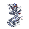
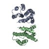
- Assembly
| Deposited unit | 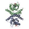
| ||||||||
|---|---|---|---|---|---|---|---|---|---|
| 1 |
| ||||||||
| 2 | 
| ||||||||
| 3 | 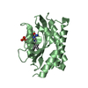
| ||||||||
| Unit cell |
|
-Components
| #1: Protein | Mass: 19531.078 Da / Num. of mol.: 2 Source method: isolated from a genetically manipulated source Source: (gene. exp.) Rhodobacter sphaeroides (bacteria) / Strain: ATCC 17025 / Gene: Rhodobacter sphaeroides / Plasmid: pET28a / Production host: #2: Chemical | ChemComp-FMN / |   Mass: 456.344 Da / Num. of mol.: 1 / Source method: obtained synthetically / Formula: C17H21N4O9P Mass: 456.344 Da / Num. of mol.: 1 / Source method: obtained synthetically / Formula: C17H21N4O9P#3: Chemical | ChemComp-FNR / | 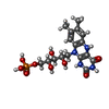  Mass: 458.360 Da / Num. of mol.: 1 / Source method: obtained synthetically / Formula: C17H23N4O9P Mass: 458.360 Da / Num. of mol.: 1 / Source method: obtained synthetically / Formula: C17H23N4O9P#4: Water | ChemComp-HOH / |  Mass: 18.015 Da / Num. of mol.: 75 / Source method: isolated from a natural source / Formula: H2O Mass: 18.015 Da / Num. of mol.: 75 / Source method: isolated from a natural source / Formula: H2O |
|---|
-Experimental details
-Experiment
| Experiment | Method: X-RAY DIFFRACTION / Number of used crystals: 3 |
|---|
- Sample preparation
| Crystal | Density Matthews: 3.78 Å3/Da / Density % sol: 67.43 % |
|---|---|
| Crystal grow | Temperature: 290 K / Method: vapor diffusion, hanging drop / pH: 7.5 Details: 0.1 M HEPES, 4.3 M NaCl, pH 7.5, VAPOR DIFFUSION, HANGING DROP, temperature 290K |
-Data collection
| Diffraction | Mean temperature: 100 K |
|---|---|
| Diffraction source | Source: SYNCHROTRON / Site: CHESS  / Beamline: A1 / Wavelength: 0.987 Å / Beamline: A1 / Wavelength: 0.987 Å |
| Detector | Type: ADSC QUANTUM 210 / Detector: CCD / Date: May 19, 2011 |
| Radiation | Protocol: SINGLE WAVELENGTH / Monochromatic (M) / Laue (L): M / Scattering type: x-ray |
| Radiation wavelength | Wavelength: 0.987 Å / Relative weight: 1 |
| Reflection | Resolution: 2.34→38.956 Å / Num. obs: 21851 / % possible obs: 88.7 % / Redundancy: 3.6 % |
| Reflection shell | Resolution: 2.34→2.38 Å / % possible all: 93.1 |
- Processing
| Software |
| ||||||||||||||||
|---|---|---|---|---|---|---|---|---|---|---|---|---|---|---|---|---|---|
| Refinement | Method to determine structure: MOLECULAR REPLACEMENT / Resolution: 2.34→38.956 Å
| ||||||||||||||||
| Refinement step | Cycle: LAST / Resolution: 2.34→38.956 Å
|
