 Movie
Movie Controller

[English] 日本語
 Yorodumi
- PDB-4hks: Calcium release-activated calcium (CRAC) channel ORAI, K163W mutant -
+ Open data

- Basic information
| Entry | Database: PDB / ID: 4hks | ||||||
|---|---|---|---|---|---|---|---|
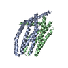
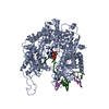
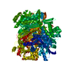
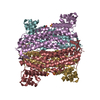
| Title | Calcium release-activated calcium (CRAC) channel ORAI, K163W mutant | ||||||
 Components Components | Calcium release-activated calcium channel protein 1 | ||||||
 Keywords Keywords | TRANSPORT PROTEIN / calcium channel / Orai1 / eukaryotic membrane protein / membrane protein / ion channel / STIM / membrane | ||||||
| Function / homology |  Function and homology information Function and homology informationstore-operated calcium entry / store-operated calcium channel activity / positive regulation of calcineurin-NFAT signaling cascade / positive regulation of calcium ion transport / calcium channel complex / calcium channel regulator activity / calcium ion transmembrane transport / calcium channel activity / membrane / identical protein binding / plasma membrane Similarity search - Function | ||||||
| Biological species |  | ||||||
| Method |  X-RAY DIFFRACTION / SYNCHROTRON / MIRAS / Resolution: 3.3516 Å X-RAY DIFFRACTION / SYNCHROTRON / MIRAS / Resolution: 3.3516 Å | ||||||
 Authors Authors | Long, S.B. / Hou, X. | ||||||
 Citation Citation | Journal: Science / Year: 2012 Title: Crystal structure of the calcium release-activated calcium channel Orai. Authors: Hou, X. / Pedi, L. / Diver, M.M. / Long, S.B. | ||||||
| History |
|
- Structure visualization
| Structure viewer | Molecule: MolmilJmol/JSmol |
|---|
- Downloads & links
-Download
| PDBx/mmCIF format | 4hks.cif.gz | 72.9 KB | Display | PDBx/mmCIF format |
|---|---|---|---|---|
| PDB format | pdb4hks.ent.gz | 53.5 KB | Display | PDB format |
| PDBx/mmJSON format | 4hks.json.gz | Tree view | PDBx/mmJSON format | |
| Others |  Other downloads Other downloads |
-Validation report
| Arichive directory | https://data.pdbj.org/pub/pdb/validation_reports/hk/4hksftp://data.pdbj.org/pub/pdb/validation_reports/hk/4hks | HTTPS FTP |
|---|
-Related structure data


-Links
 PDBj
PDBj- Assembly
| Deposited unit | 
| |||||||||
|---|---|---|---|---|---|---|---|---|---|---|
| 1 | 
| |||||||||
| Unit cell |
| |||||||||
| Components on special symmetry positions |
| |||||||||
| Details | The biological assembly is a hexamer generated from the dimer in the asymmetric unit by the three fold crystallographic axis |
-Components
| #1: Protein | Mass: 24404.424 Da / Num. of mol.: 2 / Fragment: UNP Residues 133-341 / Mutation: K163W, C224S, C283T, P276R, P277R Source method: isolated from a genetically manipulated source Source: (gene. exp.)  Pichia pastoris (fungus) / References: UniProt: Q9U6B8 Pichia pastoris (fungus) / References: UniProt: Q9U6B8#2: Chemical | ChemComp-CA / |   Mass: 40.078 Da / Num. of mol.: 1 / Source method: obtained synthetically / Formula: Ca Mass: 40.078 Da / Num. of mol.: 1 / Source method: obtained synthetically / Formula: Ca#3: Chemical | ChemComp-ZN / |   Mass: 65.409 Da / Num. of mol.: 1 / Source method: obtained synthetically / Formula: Zn Mass: 65.409 Da / Num. of mol.: 1 / Source method: obtained synthetically / Formula: Zn |
|---|
-Experimental details
-Experiment
| Experiment | Method: X-RAY DIFFRACTION / Number of used crystals: 1 |
|---|
- Sample preparation
| Crystal | Density Matthews: 2.83 Å3/Da / Density % sol: 56.5 % |
|---|---|
| Crystal grow | Temperature: 290 K / Method: vapor diffusion, hanging drop / pH: 6.5 Details: 26 % PEG 400, 1 M Ammonium Formate, 100 mM MES pH 6.5, 2 mM Calcium chloride. The crystal was soaked in: 50 % PEG 400, 1 M Ammonium Formate, 100 mM MES pH 6.5, 50 mM Calcium chloride, VAPOR ...Details: 26 % PEG 400, 1 M Ammonium Formate, 100 mM MES pH 6.5, 2 mM Calcium chloride. The crystal was soaked in: 50 % PEG 400, 1 M Ammonium Formate, 100 mM MES pH 6.5, 50 mM Calcium chloride, VAPOR DIFFUSION, HANGING DROP, temperature 290K |
-Data collection
| Diffraction | Mean temperature: 100 K |
|---|---|
| Diffraction source | Source: SYNCHROTRON / Site: NSLS  / Beamline: X29A / Wavelength: 1.075 Å / Beamline: X29A / Wavelength: 1.075 Å |
| Detector | Type: ADSC QUANTUM 315r / Detector: CCD |
| Radiation | Protocol: SINGLE WAVELENGTH / Monochromatic (M) / Laue (L): M / Scattering type: x-ray |
| Radiation wavelength | Wavelength: 1.075 Å / Relative weight: 1 |
| Reflection | Resolution: 3.35→40 Å / Num. all: 8157 / Num. obs: 8157 / % possible obs: 99.7 % / Observed criterion σ(I): -3 / Redundancy: 24.3 % / Biso Wilson estimate: 165.79 Å2 / Rsym value: 0.083 / Net I/σ(I): 35.3 |
| Reflection shell | Resolution: 3.35→3.41 Å / Redundancy: 23.4 % / Mean I/σ(I) obs: 1.3 / % possible all: 100 |
- Processing
| Software |
| ||||||||||||||||||||||||||||
|---|---|---|---|---|---|---|---|---|---|---|---|---|---|---|---|---|---|---|---|---|---|---|---|---|---|---|---|---|---|
| Refinement | Method to determine structure: MIRAS / Resolution: 3.3516→20 Å / SU ML: 0.38 / Isotropic thermal model: isotropic / σ(F): 0 / Phase error: 47.63 / Stereochemistry target values: ENGH & HUBER
| ||||||||||||||||||||||||||||
| Solvent computation | Shrinkage radii: 0.8 Å / VDW probe radii: 1.2 Å / Solvent model: FLAT BULK SOLVENT MODEL | ||||||||||||||||||||||||||||
| Displacement parameters | Biso mean: 172.8 Å2 | ||||||||||||||||||||||||||||
| Refinement step | Cycle: LAST / Resolution: 3.3516→20 Å
| ||||||||||||||||||||||||||||
| Refine LS restraints |
| ||||||||||||||||||||||||||||
| LS refinement shell |
|
