 Movie
Movie Controller

[English] 日本語
 Yorodumi
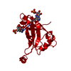
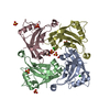
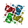
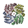
- PDB-4hi5: crystal structure of F37A mutant of borna disease virus matrix protein -
+ Open data

- Basic information
| Entry | Database: PDB / ID: 4hi5 | ||||||
|---|---|---|---|---|---|---|---|
| Title | crystal structure of F37A mutant of borna disease virus matrix protein | ||||||
 Components Components | Matrix protein | ||||||
 Keywords Keywords | VIRAL PROTEIN / VIRAL MATRIX PROTEIN / RNA BINDING / MEMBRANE BINDING / VIRUSES / SSRNA NEGATIVE-STRAND VIRUSES / MONONEGAVIRALES / BORNAVIRIDAE / BORNAVIRUS / VIRION | ||||||
| Function / homology |  Function and homology information Function and homology informationvirion component / structural constituent of virion / host cell cytoplasm / host cell plasma membrane / identical protein binding Similarity search - Function | ||||||
| Biological species |  Borna disease virus Borna disease virus | ||||||
| Method |  X-RAY DIFFRACTION / SYNCHROTRON / MOLECULAR REPLACEMENT / Resolution: 3.6 Å X-RAY DIFFRACTION / SYNCHROTRON / MOLECULAR REPLACEMENT / Resolution: 3.6 Å | ||||||
 Authors Authors | Dautel, P. / Kolenko, P. / Stubbs, M.T. | ||||||
 Citation Citation | Journal: To be Published Title: Matrix protein variants provide support for alternative borna disease virus infection pathway Authors: Dautel, P. / Kolenko, P. / Stubbs, M.T. | ||||||
| History |
|
- Structure visualization
| Structure viewer | Molecule: MolmilJmol/JSmol |
|---|
- Downloads & links
-Download
| PDBx/mmCIF format | 4hi5.cif.gz | 41.1 KB | Display | PDBx/mmCIF format |
|---|---|---|---|---|
| PDB format | pdb4hi5.ent.gz | 28.9 KB | Display | PDB format |
| PDBx/mmJSON format | 4hi5.json.gz | Tree view | PDBx/mmJSON format | |
| Others |  Other downloads Other downloads |
-Validation report
| Arichive directory | https://data.pdbj.org/pub/pdb/validation_reports/hi/4hi5ftp://data.pdbj.org/pub/pdb/validation_reports/hi/4hi5 | HTTPS FTP |
|---|
-Related structure data
| Related structure data |  4hitC  4hiuC  4hiwC  4hiyC  3f1jS S: Starting model for refinement C: citing same article ( |
|---|---|
| Similar structure data |


-Links
 PDBj
PDBj- Assembly
| Deposited unit | 
| ||||||||
|---|---|---|---|---|---|---|---|---|---|
| 1 | 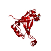
| ||||||||
| Unit cell |
|
-Components
| #1: Protein | Mass: 16201.722 Da / Num. of mol.: 1 / Mutation: F37A Source method: isolated from a genetically manipulated source Source: (gene. exp.) Borna disease virus / Gene: M / Plasmid: PMAL-C2 / Production host:  |
|---|---|
| #2: Chemical | ChemComp-C5P /   Mass: 323.197 Da / Num. of mol.: 1 / Source method: obtained synthetically / Formula: C9H14N3O8P Mass: 323.197 Da / Num. of mol.: 1 / Source method: obtained synthetically / Formula: C9H14N3O8P |
-Experimental details
-Experiment
| Experiment | Method: X-RAY DIFFRACTION / Number of used crystals: 1 |
|---|
- Sample preparation
| Crystal | Density Matthews: 3.5 Å3/Da / Density % sol: 64.87 % |
|---|---|
| Crystal grow | Temperature: 293 K / Method: vapor diffusion, hanging drop / pH: 7.1 Details: 0.2 M MgCl2, 30% w/v PEG400, 0.1 M HEPES, pH 7.1, VAPOR DIFFUSION, HANGING DROP, temperature 293K |
-Data collection
| Diffraction | Mean temperature: 100 K |
|---|---|
| Diffraction source | Source: SYNCHROTRON / Site: BESSY  / Beamline: 14.1 / Wavelength: 0.91841 / Beamline: 14.1 / Wavelength: 0.91841 |
| Detector | Type: MARMOSAIC 225 mm CCD / Detector: CCD / Date: Jul 10, 2010 / Details: MIRRORS |
| Radiation | Monochromator: SI 111 CHANNEL / Protocol: SINGLE WAVELENGTH / Monochromatic (M) / Laue (L): M / Scattering type: x-ray |
| Radiation wavelength | Wavelength: 0.91841 Å / Relative weight: 1 |
| Reflection | Resolution: 3.6→70 Å / Num. obs: 2918 / % possible obs: 100 % / Observed criterion σ(I): -3.7 / Redundancy: 11 % / Rmerge(I) obs: 0.134 / Net I/σ(I): 13.4 |
- Processing
| Software |
| ||||||||||||||||||||||||||||||||||||||||||||||||||||||||||||||||||||||||||||||||||||||||||||||||||||||||||||||||||||||||||||||||||||||||||||||||||||||||||||||||||||||||||
|---|---|---|---|---|---|---|---|---|---|---|---|---|---|---|---|---|---|---|---|---|---|---|---|---|---|---|---|---|---|---|---|---|---|---|---|---|---|---|---|---|---|---|---|---|---|---|---|---|---|---|---|---|---|---|---|---|---|---|---|---|---|---|---|---|---|---|---|---|---|---|---|---|---|---|---|---|---|---|---|---|---|---|---|---|---|---|---|---|---|---|---|---|---|---|---|---|---|---|---|---|---|---|---|---|---|---|---|---|---|---|---|---|---|---|---|---|---|---|---|---|---|---|---|---|---|---|---|---|---|---|---|---|---|---|---|---|---|---|---|---|---|---|---|---|---|---|---|---|---|---|---|---|---|---|---|---|---|---|---|---|---|---|---|---|---|---|---|---|---|---|---|
| Refinement | Method to determine structure: MOLECULAR REPLACEMENT Starting model: PDB ENTRY 3F1J Resolution: 3.6→70 Å / Cor.coef. Fo:Fc: 0.901 / Cor.coef. Fo:Fc free: 0.87 / Isotropic thermal model: Isotropic / Cross valid method: THROUGHOUT / ESU R Free: 0.775 / Stereochemistry target values: MAXIMUM LIKELIHOOD Details: HYDROGENS HAVE BEEN ADDED IN THE RIDING POSITIONS. STRUCTURE SOLUTION AND REFINEMENT WERE PERFORMED USING FREE R FOR CROSS-VALIDATION THROUGHOUT: WORKING R = 0.2352; FREE R = 0.3125 FROM A ...Details: HYDROGENS HAVE BEEN ADDED IN THE RIDING POSITIONS. STRUCTURE SOLUTION AND REFINEMENT WERE PERFORMED USING FREE R FOR CROSS-VALIDATION THROUGHOUT: WORKING R = 0.2352; FREE R = 0.3125 FROM A RANDOM TEST SET COMPRISING 10% OF ALL REFLECTIONS (273 TOTAL). FINAL REFINEMENT WAS PERFORMED USING ALL REFLECTIONS.
| ||||||||||||||||||||||||||||||||||||||||||||||||||||||||||||||||||||||||||||||||||||||||||||||||||||||||||||||||||||||||||||||||||||||||||||||||||||||||||||||||||||||||||
| Solvent computation | Ion probe radii: 0.8 Å / Shrinkage radii: 0.8 Å / VDW probe radii: 1.4 Å / Solvent model: MASK | ||||||||||||||||||||||||||||||||||||||||||||||||||||||||||||||||||||||||||||||||||||||||||||||||||||||||||||||||||||||||||||||||||||||||||||||||||||||||||||||||||||||||||
| Displacement parameters | Biso mean: 105.14 Å2 | ||||||||||||||||||||||||||||||||||||||||||||||||||||||||||||||||||||||||||||||||||||||||||||||||||||||||||||||||||||||||||||||||||||||||||||||||||||||||||||||||||||||||||
| Refinement step | Cycle: LAST / Resolution: 3.6→70 Å
| ||||||||||||||||||||||||||||||||||||||||||||||||||||||||||||||||||||||||||||||||||||||||||||||||||||||||||||||||||||||||||||||||||||||||||||||||||||||||||||||||||||||||||
| Refine LS restraints |
| ||||||||||||||||||||||||||||||||||||||||||||||||||||||||||||||||||||||||||||||||||||||||||||||||||||||||||||||||||||||||||||||||||||||||||||||||||||||||||||||||||||||||||
| LS refinement shell | Resolution: 3.601→3.694 Å / Total num. of bins used: 20 /
|
