Mass: 18.015 Da / Num. of mol.: 220 / Source method: isolated from a natural source / Formula: H2O
-
Experimental details
-
Experiment
Experiment
Method: X-RAY DIFFRACTION / Number of used crystals: 1
-
Sample preparation
Crystal
Density Matthews: 1.99 Å3/Da / Density % sol: 38 %
Crystal grow
Temperature: 290 K / Method: vapor diffusion, sitting drop / pH: 7.5 Details: EMBio JCSG+ B4: 10% PEG 8000, 8% Ethylene glycol, 100mM HEPES pH 7.5; MytuD.18400.a.A1.PS01434 at 12.8mg/ml + 2.5mM AMPPNP and MgCl2, cryo: 20% EG; the structure was solved via iodide SAD: a ...Details: EMBio JCSG+ B4: 10% PEG 8000, 8% Ethylene glycol, 100mM HEPES pH 7.5; MytuD.18400.a.A1.PS01434 at 12.8mg/ml + 2.5mM AMPPNP and MgCl2, cryo: 20% EG; the structure was solved via iodide SAD: a crystal was soaked in reservoir plus 20% ethylene glycol and 750mM NaI, anomalous amplitudes are deposited, VAPOR DIFFUSION, SITTING DROP, temperature 290K
D res high: 2.4 Å / D res low: 43.38 Å / FOM : 0.336 / FOM acentric: 0.374 / FOM centric: 0.12 / Reflection: 14132 / Reflection acentric: 12017 / Reflection centric: 2102
-
Processing
Software
Name
Version
Classification
NB
XSCALE
datascaling
PHASER
2.3.0
phasing
REFMAC
5.7.0029
refinement
PDB_EXTRACT
3.11
dataextraction
StructureStudio
datacollection
Refinement
Method to determine structure: SAD Starting model: de novo phasing with iodide ions Resolution: 1.8→38.39 Å / Cor.coef. Fo:Fc: 0.956 / Cor.coef. Fo:Fc free: 0.94 / Occupancy max: 1 / Occupancy min: 0.4 / SU B: 5.611 / SU ML: 0.087 / Isotropic thermal model: isotropic, TLS / Cross valid method: THROUGHOUT / σ(F): 0 / ESU R: 0.131 / ESU R Free: 0.126 / Stereochemistry target values: MAXIMUM LIKELIHOOD Details: HYDROGENS HAVE BEEN ADDED IN THE RIDING POSITIONS U VALUES : WITH TLS ADDED
Rfactor
Num. reflection
% reflection
Selection details
Rfree
0.2277
1645
5 %
RANDOM
Rwork
0.1884
-
-
-
all
0.1904
33216
-
-
obs
0.1904
31227
99.15 %
-
Solvent computation
Ion probe radii: 0.8 Å / Shrinkage radii: 0.8 Å / VDW probe radii: 1.2 Å / Solvent model: MASK
In the structure databanks used in Yorodumi, some data are registered as the other names, "COVID-19 virus" and "2019-nCoV". Here are the details of the virus and the list of structure data.
Jan 31, 2019. EMDB accession codes are about to change! (news from PDBe EMDB page)
EMDB accession codes are about to change! (news from PDBe EMDB page)
The allocation of 4 digits for EMDB accession codes will soon come to an end. Whilst these codes will remain in use, new EMDB accession codes will include an additional digit and will expand incrementally as the available range of codes is exhausted. The current 4-digit format prefixed with “EMD-” (i.e. EMD-XXXX) will advance to a 5-digit format (i.e. EMD-XXXXX), and so on. It is currently estimated that the 4-digit codes will be depleted around Spring 2019, at which point the 5-digit format will come into force.
The EM Navigator/Yorodumi systems omit the EMD- prefix.
Related info.:Q: What is EMD? / ID/Accession-code notation in Yorodumi/EM Navigator
Yorodumi is a browser for structure data from EMDB, PDB, SASBDB, etc.
This page is also the successor to EM Navigator detail page, and also detail information page/front-end page for Omokage search.
The word "yorodu" (or yorozu) is an old Japanese word meaning "ten thousand". "mi" (miru) is to see.
Related info.:EMDB / PDB / SASBDB / Comparison of 3 databanks / Yorodumi Search / Aug 31, 2016. New EM Navigator & Yorodumi / Yorodumi Papers / Jmol/JSmol / Function and homology information / Changes in new EM Navigator and Yorodumi
 Movie
Movie Controller

 Yorodumi
 Open data
 Basic information
 Components
 Keywords
 Function and homology information
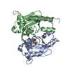
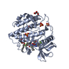
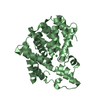
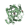
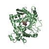
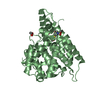
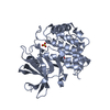
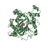
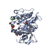
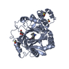
 Mycobacterium tuberculosis (bacteria)
 X-RAY DIFFRACTION /
  Authors
 Citation
 Structure visualization
 Downloads & links
 Other downloads
 PDBj
PDBj Assembly
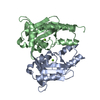

 Mass: 24.305 Da / Num. of mol.: 2 / Source method: obtained synthetically / Formula: Mg
 Mass: 18.015 Da / Num. of mol.: 220 / Source method: isolated from a natural source / Formula: H2O
 Sample preparation
 Processing