 Movie
Movie Controller


+ Open data

- Basic information
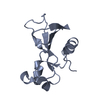
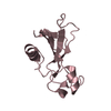
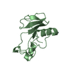
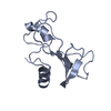
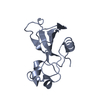
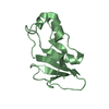
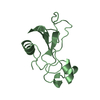
| Entry | Database: PDB / ID: 4haa | ||||||
|---|---|---|---|---|---|---|---|
| Title | Structure of Ribonuclease Binase Glu43Ala/Phe81Ala Mutant | ||||||
 Components Components | Ribonuclease | ||||||
 Keywords Keywords | HYDROLASE / endoribonuclease | ||||||
| Function / homology |  Function and homology information Function and homology informationHydrolases; Acting on ester bonds; Endoribonucleases producing 3'-phosphomonoesters / RNA endonuclease activity / hydrolase activity / RNA binding / extracellular region Similarity search - Function | ||||||
| Biological species |  | ||||||
| Method |  X-RAY DIFFRACTION / SYNCHROTRON / MOLECULAR REPLACEMENT / Resolution: 1.9 Å X-RAY DIFFRACTION / SYNCHROTRON / MOLECULAR REPLACEMENT / Resolution: 1.9 Å | ||||||
 Authors Authors | Polyakov, K.M. / Trofimov, A.A. / Mitchevich, V.A. / Dorovatovskii, P.V. / Schulga, A.A. / Makarov, A.A. / Tkach, E.N. / Goncharuk, D.A. | ||||||
 Citation Citation | Journal: Acta Crystallogr.,Sect.D / Year: 2013 Title: Structure and functional studies of the ribonuclease binase Glu43Ala/Phe81Ala mutant. Authors: Mitkevich, V.A. / Schulga, A.A. / Trofimov, A.A. / Dorovatovskii, P.V. / Goncharuk, D.A. / Tkach, E.N. / Makarov, A.A. / Polyakov, K.M. | ||||||
| History |
|
- Structure visualization
| Structure viewer | Molecule: MolmilJmol/JSmol |
|---|
- Downloads & links
-Download
| PDBx/mmCIF format | 4haa.cif.gz | 98.7 KB | Display | PDBx/mmCIF format |
|---|---|---|---|---|
| PDB format | pdb4haa.ent.gz | 77.5 KB | Display | PDB format |
| PDBx/mmJSON format | 4haa.json.gz | Tree view | PDBx/mmJSON format | |
| Others |  Other downloads Other downloads |
-Validation report
| Arichive directory | https://data.pdbj.org/pub/pdb/validation_reports/ha/4haaftp://data.pdbj.org/pub/pdb/validation_reports/ha/4haa | HTTPS FTP |
|---|
-Related structure data
| Related structure data |  1govS S: Starting model for refinement |
|---|---|
| Similar structure data |


-Links
 PDBj
PDBj- Assembly
| Deposited unit | 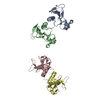
| ||||||||
|---|---|---|---|---|---|---|---|---|---|
| 1 | 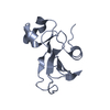
| ||||||||
| 2 | 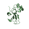
| ||||||||
| 3 | 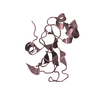
| ||||||||
| 4 | 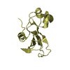
| ||||||||
| Unit cell |
|
-Components
| #1: Protein | Mass: 12093.490 Da / Num. of mol.: 4 / Fragment: UNP residues 54-162 / Mutation: E43A/F81A Source method: isolated from a genetically manipulated source Source: (gene. exp.) References: UniProt: P00649, Hydrolases; Acting on ester bonds; Endoribonucleases producing 3'-phosphomonoesters #2: Water | ChemComp-HOH / |  Mass: 18.015 Da / Num. of mol.: 264 / Source method: isolated from a natural source / Formula: H2O Mass: 18.015 Da / Num. of mol.: 264 / Source method: isolated from a natural source / Formula: H2O |
|---|
-Experimental details
-Experiment
| Experiment | Method: X-RAY DIFFRACTION / Number of used crystals: 1 |
|---|
- Sample preparation
| Crystal | Density Matthews: 2.57 Å3/Da / Density % sol: 52.15 % |
|---|---|
| Crystal grow | Temperature: 298 K / Method: vapor diffusion, hanging drop / pH: 3.5 Details: 10 mg/mL protein, reservoir: 0.1 M citric acid, pH 3.5, 3 M sodium chloride, VAPOR DIFFUSION, HANGING DROP, temperature 298K |
-Data collection
| Diffraction | Mean temperature: 100 K | |||||||||||||||
|---|---|---|---|---|---|---|---|---|---|---|---|---|---|---|---|---|
| Diffraction source | Source: SYNCHROTRON / Site: KURCHATOV SNC  / Beamline: K4.4 / Wavelength: 0.983 Å / Beamline: K4.4 / Wavelength: 0.983 Å | |||||||||||||||
| Detector | Type: MAR CCD 165 mm / Detector: CCD / Date: Nov 10, 2011 | |||||||||||||||
| Radiation | Protocol: SINGLE WAVELENGTH / Monochromatic (M) / Laue (L): M / Scattering type: x-ray | |||||||||||||||
| Radiation wavelength | Wavelength: 0.983 Å / Relative weight: 1 | |||||||||||||||
| Reflection twin |
| |||||||||||||||
| Reflection | Resolution: 1.89→60 Å / Num. all: 37967 / Num. obs: 37967 / % possible obs: 97.7 % / Observed criterion σ(F): 0 / Observed criterion σ(I): 0 / Rmerge(I) obs: 0.091 | |||||||||||||||
| Reflection shell | Resolution: 1.89→2.01 Å / Rmerge(I) obs: 0.492 / Mean I/σ(I) obs: 2.49 / Num. unique all: 5895 / % possible all: 89.9 |
- Processing
| Software |
| |||||||||||||||||||||||||||||||||||||||||||||
|---|---|---|---|---|---|---|---|---|---|---|---|---|---|---|---|---|---|---|---|---|---|---|---|---|---|---|---|---|---|---|---|---|---|---|---|---|---|---|---|---|---|---|---|---|---|---|
| Refinement | Method to determine structure: MOLECULAR REPLACEMENT Starting model: PDB ENTRY 1GOV Resolution: 1.9→18.73 Å / Cor.coef. Fo:Fc: 0.951 / Cor.coef. Fo:Fc free: 0.942 / SU B: 2.02 / SU ML: 0.064 / Cross valid method: THROUGHOUT / ESU R: 0.03 / ESU R Free: 0.027 / Stereochemistry target values: MAXIMUM LIKELIHOOD
| |||||||||||||||||||||||||||||||||||||||||||||
| Solvent computation | Ion probe radii: 0.8 Å / Shrinkage radii: 0.8 Å / VDW probe radii: 1.2 Å / Solvent model: BABINET MODEL WITH MASK | |||||||||||||||||||||||||||||||||||||||||||||
| Displacement parameters | Biso mean: 27.062 Å2
| |||||||||||||||||||||||||||||||||||||||||||||
| Refine analyze | Luzzati coordinate error free: 0.027 Å | |||||||||||||||||||||||||||||||||||||||||||||
| Refinement step | Cycle: LAST / Resolution: 1.9→18.73 Å
| |||||||||||||||||||||||||||||||||||||||||||||
| Refine LS restraints |
| |||||||||||||||||||||||||||||||||||||||||||||
| LS refinement shell | Resolution: 1.9→1.949 Å / Total num. of bins used: 20
|
