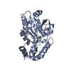
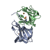
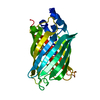
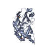
- PDB-4h7l: Crystal structure of Plim_4148 protein from Planctomyces limnophilus -
+
Open data
ID or keywords:
Loading...
-
Basic information
Entry
Database: PDB / ID: 4h7l
Title
Crystal structure of Plim_4148 protein from Planctomyces limnophilus
Components
uncharacterized protein
Keywords
Structural Genomics / Unknown Function / PSI-Biology / Midwest Center for Structural Genomics / MCSG / cupin
Function / homology
RmlC-like cupin domain superfamily / Jelly Rolls / RmlC-like jelly roll fold / Jelly Rolls / Sandwich / Mainly Beta / metal ion binding / COPPER (II) ION / Cupin 2 conserved barrel domain protein
Function and homology information
Biological species
Planctomyces limnophilus (bacteria)
Method
X-RAY DIFFRACTION / SYNCHROTRON / SAD / Resolution: 2.452 Å
Monochromator: double crystal / Protocol: SINGLE WAVELENGTH / Monochromatic (M) / Laue (L): M / Scattering type: x-ray
Radiation wavelength
Wavelength: 0.97931 Å / Relative weight: 1
Reflection
Resolution: 2.45→30 Å / Num. all: 10974 / Num. obs: 10779 / % possible obs: 98.2 % / Observed criterion σ(I): -3 / Redundancy: 4.7 % / Biso Wilson estimate: 60.1 Å2 / Rmerge(I) obs: 0.093 / Net I/σ(I): 17.93
Reflection shell
Resolution: 2.45→2.49 Å / Redundancy: 4.8 % / Rmerge(I) obs: 0.763 / Mean I/σ(I) obs: 2.18 / Num. unique all: 549 / % possible all: 99.8
-
Processing
Software
Name
Version
Classification
SBC-Collect
datacollection
SHELX
modelbuilding
MLPHARE
phasing
DM
modelbuilding
BUCCANEER
modelbuilding
ARP/wARP
modelbuilding
Coot
modelbuilding
PHENIX
(phenix.refine: dev_1096)
refinement
HKL-3000
datareduction
HKL-3000
datascaling
SHELX
phasing
DM
phasing
BUCCANEER
phasing
Refinement
Method to determine structure: SAD / Resolution: 2.452→29.051 Å / SU ML: 0.27 / Isotropic thermal model: isotropic / Cross valid method: THROUGHOUT / σ(F): 0 / Phase error: 23.54 / Stereochemistry target values: ML / Details: HYDROGEN ATOMS HAVE BEEN ADDED AT RIDING POSITIONS
Rfactor
Num. reflection
% reflection
Selection details
Rfree
0.2257
810
7.52 %
random
Rwork
0.1766
-
-
-
all
0.1804
10775
-
-
obs
0.1804
10775
98.22 %
-
Solvent computation
Shrinkage radii: 0.9 Å / VDW probe radii: 1.11 Å / Solvent model: FLAT BULK SOLVENT MODEL
Refinement step
Cycle: LAST / Resolution: 2.452→29.051 Å
Protein
Nucleic acid
Ligand
Solvent
Total
Num. atoms
1802
0
2
11
1815
Refine LS restraints
Refine-ID
Type
Dev ideal
Number
X-RAY DIFFRACTION
f_bond_d
0.014
1862
X-RAY DIFFRACTION
f_angle_d
0.853
2549
X-RAY DIFFRACTION
f_dihedral_angle_d
15.311
653
X-RAY DIFFRACTION
f_chiral_restr
0.049
288
X-RAY DIFFRACTION
f_plane_restr
0.003
327
LS refinement shell
Resolution (Å)
Rfactor Rfree
Num. reflection Rfree
Rfactor Rwork
Num. reflection Rwork
Refine-ID
% reflection obs (%)
2.452-2.6055
0.2903
135
0.2431
1625
X-RAY DIFFRACTION
98
2.6055-2.8065
0.3014
139
0.248
1645
X-RAY DIFFRACTION
99
2.8065-3.0887
0.3128
144
0.2321
1637
X-RAY DIFFRACTION
99
3.0887-3.5349
0.2361
138
0.1934
1665
X-RAY DIFFRACTION
99
3.5349-4.4511
0.2383
130
0.1536
1670
X-RAY DIFFRACTION
98
4.4511-29.0533
0.1573
124
0.1492
1723
X-RAY DIFFRACTION
96
Refinement TLS params.
Method: refined / Refine-ID: X-RAY DIFFRACTION
ID
L11 (°2)
L12 (°2)
L13 (°2)
L22 (°2)
L23 (°2)
L33 (°2)
S11 (Å °)
S12 (Å °)
S13 (Å °)
S21 (Å °)
S22 (Å °)
S23 (Å °)
S31 (Å °)
S32 (Å °)
S33 (Å °)
T11 (Å2)
T12 (Å2)
T13 (Å2)
T22 (Å2)
T23 (Å2)
T33 (Å2)
Origin x (Å)
Origin y (Å)
Origin z (Å)
1
5.0908
-0.8849
3.4447
3.7953
-0.594
7.3635
0.288
0.0191
-0.5004
-0.373
0.1722
0.5354
0.825
0.0112
-0.4358
0.6015
0.0297
-0.0594
0.3381
-0.049
0.6001
3.6451
48.7391
13.7635
2
3.0081
-1.2325
1.1643
1.5037
-0.4023
3.3736
0.2524
0.0517
-0.4141
-0.1723
0.0971
0.461
0.4979
-0.1101
-0.3433
0.5542
0.0709
-0.0719
0.3615
0.014
0.6607
-9.4231
57.7034
8.0946
3
2.3589
-0.5265
1.0248
4.2438
0.4186
2.1845
-0.219
-0.1622
0.6082
0.0429
0.2834
-0.0263
-0.5281
-0.0797
-0.0062
0.7405
0.0825
-0.0951
0.4307
-0.0249
0.5993
-2.3261
72.4052
14.6805
4
8.2499
-0.2835
1.3694
1.4877
-1.9453
3.7169
0.2646
-0.7575
0.527
0.4514
0.1739
0.3918
-0.6582
-0.0515
-0.4877
0.7222
0.1043
0.0577
0.4446
-0.0278
0.6323
1.8161
69.5455
18.5079
5
2.6777
-0.4915
2.9173
0.9139
0.7511
5.9388
0.2977
0.0555
0.3144
-0.2757
-0.1361
-0.0928
-0.6379
0.5003
-0.2815
0.6
-0.0556
0.0718
0.3955
-0.0723
0.5602
13.4742
66.2376
27.4457
6
3.5219
0.3496
-3.8141
5.1384
0.8314
4.4117
-0.2475
-0.177
-0.1026
-0.6727
-0.8886
0.0052
0.0538
1.5245
0.8236
0.5176
0.0402
-0.1007
0.617
0.118
0.5647
9.6628
61.0459
14.9594
7
2.2759
0.6403
1.2133
2.8519
0.3125
5.2578
0.0135
0.0291
0.1889
-0.1586
-0.0272
-0.2707
-0.2546
0.5431
0.0227
0.4
0.0318
0.0123
0.3106
0.0048
0.5227
13.8691
61.8105
21.4969
Refinement TLS group
ID
Refine-ID
Refine TLS-ID
Selection details
1
X-RAY DIFFRACTION
1
chain 'A' and (resid10through44 )
2
X-RAY DIFFRACTION
2
chain 'A' and (resid45through124 )
3
X-RAY DIFFRACTION
3
chain 'B' and (resid11through29 )
4
X-RAY DIFFRACTION
4
chain 'B' and (resid30through51 )
5
X-RAY DIFFRACTION
5
chain 'B' and (resid52through63 )
6
X-RAY DIFFRACTION
6
chain 'B' and (resid64through72 )
7
X-RAY DIFFRACTION
7
chain 'B' and (resid73through127 )
+
About Yorodumi
-
News
-
Feb 9, 2022. New format data for meta-information of EMDB entries
New format data for meta-information of EMDB entries
Version 3 of the EMDB header file is now the official format.
The previous official version 1.9 will be removed from the archive.
In the structure databanks used in Yorodumi, some data are registered as the other names, "COVID-19 virus" and "2019-nCoV". Here are the details of the virus and the list of structure data.
Jan 31, 2019. EMDB accession codes are about to change! (news from PDBe EMDB page)
EMDB accession codes are about to change! (news from PDBe EMDB page)
The allocation of 4 digits for EMDB accession codes will soon come to an end. Whilst these codes will remain in use, new EMDB accession codes will include an additional digit and will expand incrementally as the available range of codes is exhausted. The current 4-digit format prefixed with “EMD-” (i.e. EMD-XXXX) will advance to a 5-digit format (i.e. EMD-XXXXX), and so on. It is currently estimated that the 4-digit codes will be depleted around Spring 2019, at which point the 5-digit format will come into force.
The EM Navigator/Yorodumi systems omit the EMD- prefix.
Related info.:Q: What is EMD? / ID/Accession-code notation in Yorodumi/EM Navigator
Yorodumi is a browser for structure data from EMDB, PDB, SASBDB, etc.
This page is also the successor to EM Navigator detail page, and also detail information page/front-end page for Omokage search.
The word "yorodu" (or yorozu) is an old Japanese word meaning "ten thousand". "mi" (miru) is to see.
Related info.:EMDB / PDB / SASBDB / Comparison of 3 databanks / Yorodumi Search / Aug 31, 2016. New EM Navigator & Yorodumi / Yorodumi Papers / Jmol/JSmol / Function and homology information / Changes in new EM Navigator and Yorodumi
 Movie
Movie Controller

 Yorodumi
 Open data
 Basic information
 Components
 Keywords
 Function and homology information
 Planctomyces limnophilus (bacteria)
 X-RAY DIFFRACTION /
  Authors
 Structure visualization
 Downloads & links
 Other downloads
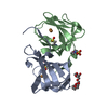


 PDBj
PDBj Assembly

 Mass: 63.546 Da / Num. of mol.: 2 / Source method: obtained synthetically / Formula: Cu
 Mass: 18.015 Da / Num. of mol.: 11 / Source method: isolated from a natural source / Formula: H2O
 Sample preparation
 / Beamline: 19-ID / Wavelength: 0.97931 Å
 Processing