- PDB-4h4j: Crystal structure of a N-acetylmuramoyl-L-alanine amidase (BACUNI... -
+
Open data
ID or keywords:
Loading...
-
Basic information
Entry
Database: PDB / ID: 4h4j
Title
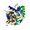
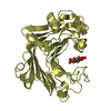
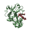
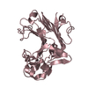
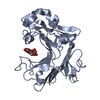
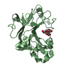
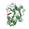
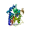
Crystal structure of a N-acetylmuramoyl-L-alanine amidase (BACUNI_02947) from Bacteroides uniformis ATCC 8492 at 1.15 A resolution
Components
hypothetical protein
Keywords
Structural Genomics / Unknown Function / PF07313 family protein / DUF 1460 / Joint Center for Structural Genomics / JCSG / Protein Structure Initiative / PSI-BIOLOGY / STRUCTURAL GENOMICS UNKNOWN FUNCTION
Mass: 18.015 Da / Num. of mol.: 431 / Source method: isolated from a natural source / Formula: H2O
Has protein modification
Y
Sequence details
THE CONSTRUCT (24-262) WAS EXPRESSED WITH A PURIFICATION TAG MGSDKIHHHHHHENLYFQG. THE TAG WAS ...THE CONSTRUCT (24-262) WAS EXPRESSED WITH A PURIFICATION TAG MGSDKIHHHHHHENLYFQG. THE TAG WAS REMOVED WITH TEV PROTEASE LEAVING ONLY A GLYCINE (0) FOLLOWED BY THE TARGET SEQUENCE.
-
Experimental details
-
Experiment
Experiment
Method: X-RAY DIFFRACTION / Number of used crystals: 1
-
Sample preparation
Crystal
Density Matthews: 2 Å3/Da / Density % sol: 38.58 %
Crystal grow
Temperature: 277 K / Method: vapor diffusion, sitting drop Details: 30.0% polyethylene glycol 1500, NANODROP, VAPOR DIFFUSION, SITTING DROP, temperature 277K
Monochromator: double crystal Si(111) / Protocol: SAD / Monochromatic (M) / Laue (L): M / Scattering type: x-ray
Radiation wavelength
Wavelength: 0.9794 Å / Relative weight: 1
Reflection
Resolution: 1.15→28.666 Å / Num. obs: 71198 / % possible obs: 92.1 % / Observed criterion σ(I): -3 / Biso Wilson estimate: 11.453 Å2 / Rmerge(I) obs: 0.067 / Net I/σ(I): 12.95
Reflection shell
Diffraction-ID: 1
Resolution (Å)
Redundancy (%)
Rmerge(I) obs
Mean I/σ(I) obs
Num. measured obs
Num. unique obs
% possible all
1.15-1.19
13.15
0.883
1.84
49534
8700
60.2
1.19-1.24
0.77
2.4
86579
13308
85.7
1.24-1.3
0.611
3.2
105067
14915
95.2
1.3-1.36
0.422
4.4
82851
12386
95.1
1.36-1.45
0.308
6.2
108285
15116
96.5
1.45-1.56
0.198
9
97412
14159
97
1.56-1.72
0.129
13.1
102725
14552
96.3
1.72-1.97
0.082
19.4
100246
14475
97.6
1.97-2.48
0.058
28.8
103841
14546
98.9
2.48-28.67
0.045
34.9
99845
14598
98.4
-
Phasing
Phasing
Method: SAD
-
Processing
Software
Name
Version
Classification
NB
MolProbity
3beta29
modelbuilding
PDB_EXTRACT
3.1
dataextraction
SHELX
phasing
SHARP
phasing
XSCALE
March15, 2012
datascaling
REFMAC
5.5.0110
refinement
XDS
datareduction
SHELXD
phasing
Refinement
Method to determine structure: SAD / Resolution: 1.15→28.666 Å / Cor.coef. Fo:Fc: 0.98 / Cor.coef. Fo:Fc free: 0.972 / Occupancy max: 1 / Occupancy min: 0.15 / SU B: 1.11 / SU ML: 0.023 / Cross valid method: THROUGHOUT / σ(F): 0 / ESU R: 0.038 / ESU R Free: 0.039 Stereochemistry target values: MAXIMUM LIKELIHOOD WITH PHASES Details: 1. A MET-INHIBITION PROTOCOL WAS USED FOR SELENOMETHIONINE INCORPORATION DURING PROTEIN EXPRESSION. THE OCCUPANCY OF THE SE ATOMS IN THE MSE RESIDUES WAS REDUCED TO 0.75 FOR THE REDUCED ...Details: 1. A MET-INHIBITION PROTOCOL WAS USED FOR SELENOMETHIONINE INCORPORATION DURING PROTEIN EXPRESSION. THE OCCUPANCY OF THE SE ATOMS IN THE MSE RESIDUES WAS REDUCED TO 0.75 FOR THE REDUCED SCATTERING POWER DUE TO PARTIAL S-MET INCORPORATION. 2. HYDROGENS HAVE BEEN ADDED IN THE RIDING POSITIONS.
Rfactor
Num. reflection
% reflection
Selection details
Rfree
0.164
3572
5 %
RANDOM
Rwork
0.1306
-
-
-
obs
0.1323
71147
91.92 %
-
Solvent computation
Ion probe radii: 0.8 Å / Shrinkage radii: 0.8 Å / VDW probe radii: 1.2 Å / Solvent model: MASK
In the structure databanks used in Yorodumi, some data are registered as the other names, "COVID-19 virus" and "2019-nCoV". Here are the details of the virus and the list of structure data.
Jan 31, 2019. EMDB accession codes are about to change! (news from PDBe EMDB page)
EMDB accession codes are about to change! (news from PDBe EMDB page)
The allocation of 4 digits for EMDB accession codes will soon come to an end. Whilst these codes will remain in use, new EMDB accession codes will include an additional digit and will expand incrementally as the available range of codes is exhausted. The current 4-digit format prefixed with “EMD-” (i.e. EMD-XXXX) will advance to a 5-digit format (i.e. EMD-XXXXX), and so on. It is currently estimated that the 4-digit codes will be depleted around Spring 2019, at which point the 5-digit format will come into force.
The EM Navigator/Yorodumi systems omit the EMD- prefix.
Related info.:Q: What is EMD? / ID/Accession-code notation in Yorodumi/EM Navigator
Yorodumi is a browser for structure data from EMDB, PDB, SASBDB, etc.
This page is also the successor to EM Navigator detail page, and also detail information page/front-end page for Omokage search.
The word "yorodu" (or yorozu) is an old Japanese word meaning "ten thousand". "mi" (miru) is to see.
Related info.:EMDB / PDB / SASBDB / Comparison of 3 databanks / Yorodumi Search / Aug 31, 2016. New EM Navigator & Yorodumi / Yorodumi Papers / Jmol/JSmol / Function and homology information / Changes in new EM Navigator and Yorodumi
 Movie
Movie Controller

 Yorodumi
 Open data
 Basic information
 Components
 Keywords
 Function and homology information
 Bacteroides uniformis (bacteria)
 X-RAY DIFFRACTION /
  Authors
 Citation
 Structure visualization
 Downloads & links
 Other downloads
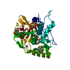


 PDBj
PDBj Assembly
 Mass: 18.015 Da / Num. of mol.: 431 / Source method: isolated from a natural source / Formula: H2O
 Sample preparation
 / Beamline: BL12-2 / Wavelength: 0.9794
  Processing