Mass: 18.015 Da / Num. of mol.: 797 / Source method: isolated from a natural source / Formula: H2O
-
Details
Has protein modification
Y
Sequence details
THE CONSTRUCT WAS EXPRESSED WITH A PURIFICATION TAG MGSDKIHHHHHHENLYFQG. THE TAG WAS REMOVED WITH ...THE CONSTRUCT WAS EXPRESSED WITH A PURIFICATION TAG MGSDKIHHHHHHENLYFQG. THE TAG WAS REMOVED WITH TEV PROTEASE LEAVING ONLY A GLYCINE (0) FOLLOWED BY RESIDUES 28-366 OF THE TARGET SEQUENCE.
-
Experimental details
-
Experiment
Experiment
Method: X-RAY DIFFRACTION / Number of used crystals: 1
-
Sample preparation
Crystal
Density Matthews: 2.18 Å3/Da / Density % sol: 43.58 %
Crystal grow
Temperature: 277 K / Method: vapor diffusion, sitting drop / pH: 6 Details: 40.0% polyethylene glycol 400, 5.0% polyethylene glycol 3000, 0.1M MES pH 6.0, NANODROP, VAPOR DIFFUSION, SITTING DROP, temperature 277K
Type: MARMOSAIC 325 mm CCD / Detector: CCD / Date: Jul 22, 2011 Details: Flat mirror (vertical focusing); single crystal Si(111) bent monochromator (ho rizontal focusing)
Radiation
Monochromator: single crystal Si(111) bent / Protocol: SAD / Monochromatic (M) / Laue (L): M / Scattering type: x-ray
Radiation wavelength
Wavelength: 0.97895 Å / Relative weight: 1
Reflection
Resolution: 1.8→29.135 Å / Num. all: 89110 / Num. obs: 89110 / % possible obs: 93.6 % / Redundancy: 12.7 % / Rsym value: 0.133 / Net I/σ(I): 12.7
Reflection shell
Diffraction-ID: 1
Resolution (Å)
Redundancy (%)
Rmerge(I) obs
Mean I/σ(I) obs
Num. measured all
Num. unique all
Rsym value
% possible all
1.8-1.85
5.3
0.595
2.1
21739
4112
0.595
60.5
1.85-1.9
5.6
0.49
2.5
27693
4965
0.49
72.3
1.9-1.95
8.5
0.476
3.6
46551
5468
0.476
82.5
1.95-2.01
12.6
0.444
5
77223
6153
0.444
94.7
2.01-2.08
13.9
0.374
6.2
87708
6311
0.374
100
2.08-2.15
14
0.306
7.5
84863
6075
0.306
100
2.15-2.23
14
0.256
8.7
82482
5905
0.256
100
2.23-2.32
14
0.229
9.9
78557
5612
0.229
100
2.32-2.43
14
0.202
11.1
76165
5435
0.202
100
2.43-2.55
14
0.176
12.4
72574
5169
0.176
100
2.55-2.68
14
0.166
13.5
69762
4969
0.166
100
2.68-2.85
14.1
0.14
15.9
65403
4652
0.14
100
2.85-3.04
14.1
0.119
18.4
61751
4385
0.119
100
3.04-3.29
14.1
0.104
21.6
57493
4075
0.104
100
3.29-3.6
14.1
0.094
24.6
53291
3781
0.094
100
3.6-4.02
14
0.087
27.7
47928
3415
0.087
100
4.02-4.65
14.1
0.083
29.6
42056
2991
0.083
100
4.65-5.69
14.1
0.09
28.8
36148
2571
0.09
100
5.69-8.05
14
0.098
27
27642
1977
0.098
100
8.05-29.135
13.6
0.088
30
14860
1089
0.088
98.1
-
Phasing
Phasing
Method: SAD
-
Processing
Software
Name
Version
Classification
NB
MolProbity
3beta29
modelbuilding
PDB_EXTRACT
3.1
dataextraction
SHELX
phasing
SHARP
phasing
SCALA
3.3.15
datascaling
BUSTER-TNT
2.10.0
refinement
MOSFLM
datareduction
SHELXD
phasing
BUSTER
2.10.0
refinement
Refinement
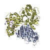
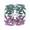
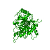
Method to determine structure: SAD / Resolution: 1.8→29.135 Å / Cor.coef. Fo:Fc: 0.9455 / Cor.coef. Fo:Fc free: 0.9373 / Occupancy max: 1 / Occupancy min: 0.33 / Cross valid method: THROUGHOUT / σ(F): 0 Details: 1. ATOM RECORD CONTAINS SUM OF TLS AND RESIDUAL B FACTORS. ANISOU RECORD CONTAINS SUM OF TLS AND RESIDUAL U FACTORS. 2. A MET-INHIBITION PROTOCOL WAS USED FOR SELENOMETHIONINE INCORPORATION ...Details: 1. ATOM RECORD CONTAINS SUM OF TLS AND RESIDUAL B FACTORS. ANISOU RECORD CONTAINS SUM OF TLS AND RESIDUAL U FACTORS. 2. A MET-INHIBITION PROTOCOL WAS USED FOR SELENOMETHIONINE INCORPORATION DURING PROTEIN EXPRESSION. THE OCCUPANCY OF THE SE ATOMS IN THE MSE RESIDUES WAS REDUCED TO 0.75 TO ACCOUNT FOR THE REDUCED SCATTERING POWER DUE TO PARTIAL S-MET INCORPORATION. 3. POLYETHYLENE GLYCOL FRAGMENTS (1PE,PE4,PG6,AND PG4) FROM THE CRYSTALLIZATION CONDITIONS AND SODIUM (NA) FROM THE PURIFICATION BUFFER HAVE BEEN MODELED IN THE SOLVENT STRUCTURE. 4. AN UNKNOWN LIGAND (UNL) WAS MODELED IN THE PUTATIVE ACTIVE SITE OF EACH PROTOMER. 5. 18 N-TERMINAL RESIDUES OF A AND C AND 11 RESIDUES OF B MOLECULES WERE NOT MODELED 6. NCS RESTRAINTS WERE APPLIED USING BUSTER'S LSSR RESTRAINT REPRESENTATION (-AUTONCS). 7. THE REFINEMENT WAS RESTRAINED AGAINST THE SAD PHASES. 8. HYDROGEN ATOMS WERE INCLUDED IN THE REFINEMENT AND TREATED BY BUSTER AS CONTRIBUTING TO FCALC.
In the structure databanks used in Yorodumi, some data are registered as the other names, "COVID-19 virus" and "2019-nCoV". Here are the details of the virus and the list of structure data.
Jan 31, 2019. EMDB accession codes are about to change! (news from PDBe EMDB page)
EMDB accession codes are about to change! (news from PDBe EMDB page)
The allocation of 4 digits for EMDB accession codes will soon come to an end. Whilst these codes will remain in use, new EMDB accession codes will include an additional digit and will expand incrementally as the available range of codes is exhausted. The current 4-digit format prefixed with “EMD-” (i.e. EMD-XXXX) will advance to a 5-digit format (i.e. EMD-XXXXX), and so on. It is currently estimated that the 4-digit codes will be depleted around Spring 2019, at which point the 5-digit format will come into force.
The EM Navigator/Yorodumi systems omit the EMD- prefix.
Related info.:Q: What is EMD? / ID/Accession-code notation in Yorodumi/EM Navigator
Yorodumi is a browser for structure data from EMDB, PDB, SASBDB, etc.
This page is also the successor to EM Navigator detail page, and also detail information page/front-end page for Omokage search.
The word "yorodu" (or yorozu) is an old Japanese word meaning "ten thousand". "mi" (miru) is to see.
Related info.:EMDB / PDB / SASBDB / Comparison of 3 databanks / Yorodumi Search / Aug 31, 2016. New EM Navigator & Yorodumi / Yorodumi Papers / Jmol/JSmol / Function and homology information / Changes in new EM Navigator and Yorodumi
 Movie
Movie Controller

 Yorodumi
 Open data
 Basic information
 Components
 Keywords
 Function and homology information
 Bacteroides thetaiotaomicron (bacteria)
 X-RAY DIFFRACTION /
  Authors
 Citation
 Structure visualization
 Downloads & links
 Other downloads
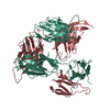


 PDBj
PDBj


 Assembly
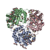


 Mass: 238.278 Da / Num. of mol.: 7 / Source method: obtained synthetically / Formula: C10H22O6 / Comment: precipitant*YM
 Mass: 354.436 Da / Num. of mol.: 2 / Source method: obtained synthetically / Formula: C16H34O8 / Comment: precipitant*YM
 Mass: 194.226 Da / Num. of mol.: 5 / Source method: obtained synthetically / Formula: C8H18O5 / Comment: precipitant*YM
 Mass: 266.331 Da / Num. of mol.: 2 / Source method: obtained synthetically / Formula: C12H26O6
 Mass: 22.990 Da / Num. of mol.: 1 / Source method: obtained synthetically / Formula: Na
 Sample preparation
 / Beamline: BL11-1 / Wavelength: 0.97895
  Processing