 Movie
Movie Controller

[English] 日本語
 Yorodumi
- PDB-4h33: Crystal structure of a voltage-gated K+ channel pore module in a ... -
+ Open data

- Basic information
| Entry | Database: PDB / ID: 4h33 | ||||||
|---|---|---|---|---|---|---|---|
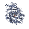
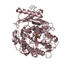
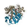
| Title | Crystal structure of a voltage-gated K+ channel pore module in a closed state in lipid membranes, tetragonal crystal form | ||||||
 Components Components | Lmo2059 protein | ||||||
 Keywords Keywords | MEMBRANE PROTEIN / BILAYERS / KVLM / LIPIDIC CUBIC PHASE (LCP) / PORE MODULE / ION CHANNEL | ||||||
| Function / homology |  Function and homology information Function and homology informationaction potential / voltage-gated potassium channel activity / voltage-gated potassium channel complex / potassium ion transmembrane transport / membrane Similarity search - Function | ||||||
| Biological species |  Listeria monocytogenes (bacteria) Listeria monocytogenes (bacteria) | ||||||
| Method |  X-RAY DIFFRACTION / SYNCHROTRON / MOLECULAR REPLACEMENT / Resolution: 3.1 Å X-RAY DIFFRACTION / SYNCHROTRON / MOLECULAR REPLACEMENT / Resolution: 3.1 Å | ||||||
 Authors Authors | Santos, J.S. / Asmar-Rovira, G.A. / Han, G.W. / Liu, W. / Syeda, R. / Cherezov, V. / Baker, K.A. / Stevens, R.C. / Montal, M. | ||||||
 Citation Citation | Journal: J.Biol.Chem. / Year: 2012 Title: Crystal Structure of a Voltage-gated K+ Channel Pore Module in a Closed State in Lipid Membranes. Authors: Santos, J.S. / Asmar-Rovira, G.A. / Han, G.W. / Liu, W. / Syeda, R. / Cherezov, V. / Baker, K.A. / Stevens, R.C. / Montal, M. | ||||||
| History |
|
- Structure visualization
| Structure viewer | Molecule: MolmilJmol/JSmol |
|---|
- Downloads & links
-Download
| PDBx/mmCIF format | 4h33.cif.gz | 52.5 KB | Display | PDBx/mmCIF format |
|---|---|---|---|---|
| PDB format | pdb4h33.ent.gz | 36.4 KB | Display | PDB format |
| PDBx/mmJSON format | 4h33.json.gz | Tree view | PDBx/mmJSON format | |
| Others |  Other downloads Other downloads |
-Validation report
| Arichive directory | https://data.pdbj.org/pub/pdb/validation_reports/h3/4h33ftp://data.pdbj.org/pub/pdb/validation_reports/h3/4h33 | HTTPS FTP |
|---|
-Related structure data
| Related structure data |  4h37C 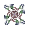 1orqS 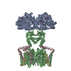 2a79S  2atkS  3effS C: citing same article ( S: Starting model for refinement |
|---|---|
| Similar structure data |


-Links
 PDBj
PDBj


- Assembly
| Deposited unit | 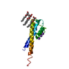
| ||||||||||||
|---|---|---|---|---|---|---|---|---|---|---|---|---|---|
| 1 | 
| ||||||||||||
| Unit cell |
| ||||||||||||
| Components on special symmetry positions |
|
-Components
| #1: Protein | Mass: 15561.433 Da / Num. of mol.: 1 Fragment: KVLM PORE MODULE, TRUNCATED C-TERMINUS (UNP RESIDUES 98-233) Mutation: A98C Source method: isolated from a genetically manipulated source Source: (gene. exp.) Listeria monocytogenes (bacteria) / Strain: ATCC BAA-679 / EGD-e / Gene: lmo2059 / Plasmid: pQE-70 / Production host: | ||
|---|---|---|---|
| #2: Chemical |   Mass: 39.098 Da / Num. of mol.: 3 / Source method: obtained synthetically / Formula: K Mass: 39.098 Da / Num. of mol.: 3 / Source method: obtained synthetically / Formula: K#3: Chemical | ChemComp-OLC / (   Mass: 356.540 Da / Num. of mol.: 4 / Source method: obtained synthetically / Formula: C21H40O4 Mass: 356.540 Da / Num. of mol.: 4 / Source method: obtained synthetically / Formula: C21H40O4 |
-Experimental details
-Experiment
| Experiment | Method: X-RAY DIFFRACTION / Number of used crystals: 32 |
|---|
- Sample preparation
| Crystal | Density Matthews: 2.47 Å3/Da / Density % sol: 50.3 % |
|---|---|
| Crystal grow | Temperature: 293 K / pH: 7 Details: 26-30% (v/v) PEG-MME-550, 0.5 M to 0.6 M ammonium sulfate, and 50 mM ADA, Lipidic Cubic Phase (LCP) , pH 7.0, temperature 293K |
-Data collection
| Diffraction | Mean temperature: 100 K |
|---|---|
| Diffraction source | Source: SYNCHROTRON / Site: APS  / Beamline: 23-ID-D / Wavelength: 1.033 Å / Beamline: 23-ID-D / Wavelength: 1.033 Å |
| Detector | Type: MARMOSAIC 300 mm CCD / Detector: CCD / Date: Mar 18, 2011 |
| Radiation | Protocol: SINGLE WAVELENGTH / Monochromatic (M) / Laue (L): M / Scattering type: x-ray |
| Radiation wavelength | Wavelength: 1.033 Å / Relative weight: 1 |
| Reflection | Resolution: 3.1→50 Å / Num. obs: 2994 / % possible obs: 94.4 % / Redundancy: 7.1 % / Biso Wilson estimate: 80.28 Å2 / Rmerge(I) obs: 0.132 / Net I/σ(I): 18.6 |
| Reflection shell | Resolution: 3.1→3.21 Å / Redundancy: 5.5 % / Rmerge(I) obs: 0.92 / Mean I/σ(I) obs: 2.45 / % possible all: 75 |
- Processing
| Software |
| ||||||||||||||||||||||||||||||||||||||||||||||||||||||||||||||||||||||||||||||||||||||||||||||||||||||||||||||||||
|---|---|---|---|---|---|---|---|---|---|---|---|---|---|---|---|---|---|---|---|---|---|---|---|---|---|---|---|---|---|---|---|---|---|---|---|---|---|---|---|---|---|---|---|---|---|---|---|---|---|---|---|---|---|---|---|---|---|---|---|---|---|---|---|---|---|---|---|---|---|---|---|---|---|---|---|---|---|---|---|---|---|---|---|---|---|---|---|---|---|---|---|---|---|---|---|---|---|---|---|---|---|---|---|---|---|---|---|---|---|---|---|---|---|---|---|
| Refinement | Method to determine structure: MOLECULAR REPLACEMENT Starting model: PDB ENTRY 3EFF, 2ATK, 2A79 and 1ORQ Resolution: 3.1→16.4 Å / Cor.coef. Fo:Fc: 0.8555 / Cor.coef. Fo:Fc free: 0.7825 / Cross valid method: THROUGHOUT / σ(F): 0
| ||||||||||||||||||||||||||||||||||||||||||||||||||||||||||||||||||||||||||||||||||||||||||||||||||||||||||||||||||
| Displacement parameters | Biso mean: 114.2 Å2
| ||||||||||||||||||||||||||||||||||||||||||||||||||||||||||||||||||||||||||||||||||||||||||||||||||||||||||||||||||
| Refine analyze | Luzzati coordinate error obs: 1.248 Å | ||||||||||||||||||||||||||||||||||||||||||||||||||||||||||||||||||||||||||||||||||||||||||||||||||||||||||||||||||
| Refinement step | Cycle: LAST / Resolution: 3.1→16.4 Å
| ||||||||||||||||||||||||||||||||||||||||||||||||||||||||||||||||||||||||||||||||||||||||||||||||||||||||||||||||||
| Refine LS restraints |
| ||||||||||||||||||||||||||||||||||||||||||||||||||||||||||||||||||||||||||||||||||||||||||||||||||||||||||||||||||
| LS refinement shell | Resolution: 3.1→3.47 Å / Total num. of bins used: 5
| ||||||||||||||||||||||||||||||||||||||||||||||||||||||||||||||||||||||||||||||||||||||||||||||||||||||||||||||||||
| Refinement TLS params. | Method: refined / Origin x: 15.3567 Å / Origin y: -5.4997 Å / Origin z: 34.4487 Å
| ||||||||||||||||||||||||||||||||||||||||||||||||||||||||||||||||||||||||||||||||||||||||||||||||||||||||||||||||||
| Refinement TLS group |
|
