 Movie
Movie Controller


+ Open data

- Basic information
| Entry | Database: PDB / ID: 4h2n | |||||||||
|---|---|---|---|---|---|---|---|---|---|---|
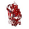
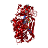
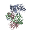
| Title | Crystal structure of MHPCO, Y270F mutant | |||||||||
 Components Components | 2-methyl-3-hydroxypyridine-5-carboxylic acid oxygenase | |||||||||
 Keywords Keywords | OXIDOREDUCTASE / FAD-binding motif / Oxygenase / FAD / 3-hydroxypyridine-5-carboxylic acid | |||||||||
| Function / homology |  Function and homology information Function and homology information3-hydroxy-2-methylpyridine-5-carboxylate monooxygenase / FAD binding / monooxygenase activity Similarity search - Function | |||||||||
| Biological species |  Rhizobium loti (bacteria) Rhizobium loti (bacteria) | |||||||||
| Method |  X-RAY DIFFRACTION / SYNCHROTRON / MOLECULAR REPLACEMENT / Resolution: 2.302 Å X-RAY DIFFRACTION / SYNCHROTRON / MOLECULAR REPLACEMENT / Resolution: 2.302 Å | |||||||||
 Authors Authors | Kobayashi, J. / Yoshida, H. / Mikami, B. / Hayashi, H. / Kamitori, S. / Yagi, T. | |||||||||
 Citation Citation | Journal: To be Published Title: Crystal structure of 2-methyl-3-hydroxypyridine-5-carboxylic acid oxygenase Authors: Kobayashi, J. / Yoshida, H. / Mikami, B. / Hayashi, H. / Kamitori, S. / Yagi, T. | |||||||||
| History |
|
- Structure visualization
| Structure viewer | Molecule: MolmilJmol/JSmol |
|---|
- Downloads & links
-Download
| PDBx/mmCIF format | 4h2n.cif.gz | 89.2 KB | Display | PDBx/mmCIF format |
|---|---|---|---|---|
| PDB format | pdb4h2n.ent.gz | 65.3 KB | Display | PDB format |
| PDBx/mmJSON format | 4h2n.json.gz | Tree view | PDBx/mmJSON format | |
| Others |  Other downloads Other downloads |
-Validation report
| Arichive directory | https://data.pdbj.org/pub/pdb/validation_reports/h2/4h2nftp://data.pdbj.org/pub/pdb/validation_reports/h2/4h2n | HTTPS FTP |
|---|
-Related structure data
| Related structure data | 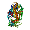 3aljC 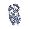 3allC 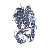 3almC  4gf7C 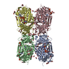 4h2pC  4jy2C  4jy3C 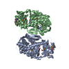 3gmbS C: citing same article ( S: Starting model for refinement |
|---|---|
| Similar structure data |


-Links
 PDBj
PDBj


- Assembly
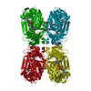
| Deposited unit | 
| ||||||||
|---|---|---|---|---|---|---|---|---|---|
| 1 | 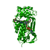
| ||||||||
| Unit cell |
|
-Components
| #1: Protein | Mass: 41782.488 Da / Num. of mol.: 1 / Mutation: Y270F Source method: isolated from a genetically manipulated source Source: (gene. exp.) Rhizobium loti (bacteria) / Strain: MAFF303099 / Gene: mlr6788 / Production host: |
|---|---|
| #2: Chemical | ChemComp-FAD /   Mass: 785.550 Da / Num. of mol.: 1 / Source method: obtained synthetically / Formula: C27H33N9O15P2 / Comment: FAD*YM Mass: 785.550 Da / Num. of mol.: 1 / Source method: obtained synthetically / Formula: C27H33N9O15P2 / Comment: FAD*YM |
| #3: Chemical | ChemComp-BME /   Mass: 78.133 Da / Num. of mol.: 1 / Source method: obtained synthetically / Formula: C2H6OS Mass: 78.133 Da / Num. of mol.: 1 / Source method: obtained synthetically / Formula: C2H6OS |
| #4: Chemical | ChemComp-GOL /   Mass: 92.094 Da / Num. of mol.: 1 / Source method: obtained synthetically / Formula: C3H8O3 Mass: 92.094 Da / Num. of mol.: 1 / Source method: obtained synthetically / Formula: C3H8O3 |
| #5: Water | ChemComp-HOH /  Mass: 18.015 Da / Num. of mol.: 94 / Source method: isolated from a natural source / Formula: H2O Mass: 18.015 Da / Num. of mol.: 94 / Source method: isolated from a natural source / Formula: H2O |
-Experimental details
-Experiment
| Experiment | Method: X-RAY DIFFRACTION / Number of used crystals: 1 |
|---|
- Sample preparation
| Crystal | Density Matthews: 2.58 Å3/Da / Density % sol: 52.33 % |
|---|---|
| Crystal grow | Temperature: 293 K / Method: vapor diffusion, hanging drop / pH: 8.5 Details: 8% PEG 8000, 0.1M Tris-HCl, pH 8.5, VAPOR DIFFUSION, HANGING DROP, temperature 293K |
-Data collection
| Diffraction | Mean temperature: 100 K |
|---|---|
| Diffraction source | Source: SYNCHROTRON / Site: Photon Factory  / Beamline: AR-NW12A / Wavelength: 1 Å / Beamline: AR-NW12A / Wavelength: 1 Å |
| Detector | Type: ADSC QUANTUM 210 / Detector: CCD / Date: May 1, 2009 |
| Radiation | Protocol: SINGLE WAVELENGTH / Monochromatic (M) / Laue (L): M / Scattering type: x-ray |
| Radiation wavelength | Wavelength: 1 Å / Relative weight: 1 |
| Reflection | Resolution: 2.3→50 Å / Num. obs: 18939 / % possible obs: 96.5 % / Redundancy: 7 % / Biso Wilson estimate: 29.31 Å2 / Rmerge(I) obs: 0.121 / Net I/σ(I): 10 |
| Reflection shell | Resolution: 2.3→2.34 Å / Redundancy: 7.3 % / Rmerge(I) obs: 0.412 / Mean I/σ(I) obs: 4.8 / Num. unique all: 968 / % possible all: 100 |
- Processing
| Software |
| ||||||||||||||||||||||||||||||||||||||||||||||||
|---|---|---|---|---|---|---|---|---|---|---|---|---|---|---|---|---|---|---|---|---|---|---|---|---|---|---|---|---|---|---|---|---|---|---|---|---|---|---|---|---|---|---|---|---|---|---|---|---|---|
| Refinement | Method to determine structure: MOLECULAR REPLACEMENT Starting model: PDB ENTRY 3GMB Resolution: 2.302→37.978 Å / Occupancy max: 1 / Occupancy min: 1 / SU ML: 0.31 / σ(F): 1.34 / Phase error: 24.6 / Stereochemistry target values: ML
| ||||||||||||||||||||||||||||||||||||||||||||||||
| Solvent computation | Shrinkage radii: 0.73 Å / VDW probe radii: 1 Å / Solvent model: FLAT BULK SOLVENT MODEL / Bsol: 19.71 Å2 / ksol: 0.32 e/Å3 | ||||||||||||||||||||||||||||||||||||||||||||||||
| Displacement parameters | Biso max: 93.96 Å2 / Biso mean: 33.5164 Å2 / Biso min: 17.87 Å2
| ||||||||||||||||||||||||||||||||||||||||||||||||
| Refinement step | Cycle: LAST / Resolution: 2.302→37.978 Å
| ||||||||||||||||||||||||||||||||||||||||||||||||
| Refine LS restraints |
| ||||||||||||||||||||||||||||||||||||||||||||||||
| LS refinement shell | Refine-ID: X-RAY DIFFRACTION / Total num. of bins used: 7
|
