 Movie
Movie Controller

[English] 日本語
 Yorodumi
- PDB-4gh5: Crystal structure of S-2-hydroxypropyl coenzyme M dehydrogenase (... -
+ Open data

- Basic information
| Entry | Database: PDB / ID: 4gh5 | ||||||
|---|---|---|---|---|---|---|---|
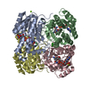
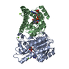
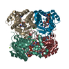
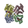
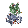
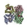
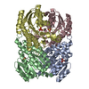
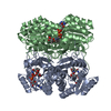
| Title | Crystal structure of S-2-hydroxypropyl coenzyme M dehydrogenase (S-HPCDH) | ||||||
 Components Components | Short-chain dehydrogenase/reductase SDR | ||||||
 Keywords Keywords | OXIDOREDUCTASE / Rossmann Fold | ||||||
| Function / homology |  Function and homology information Function and homology information2-(S)-hydroxypropyl-CoM dehydrogenase / 2-(S)-hydroxypropyl-CoM dehydrogenase activity / quinone binding / fatty acid biosynthetic process / nucleotide binding Similarity search - Function | ||||||
| Biological species |  Xanthobacter autotrophicus (bacteria) Xanthobacter autotrophicus (bacteria) | ||||||
| Method |  X-RAY DIFFRACTION / MOLECULAR REPLACEMENT / Resolution: 1.6 Å X-RAY DIFFRACTION / MOLECULAR REPLACEMENT / Resolution: 1.6 Å | ||||||
 Authors Authors | Bakelar, J.W. / Johnson, S.J. | ||||||
 Citation Citation | Journal: Arch.Biochem.Biophys. / Year: 2013 Title: Crystal structures of S-HPCDH reveal determinants of stereospecificity for R- and S-hydroxypropyl-coenzyme M dehydrogenases. Authors: Bakelar, J.W. / Sliwa, D.A. / Johnson, S.J. | ||||||
| History |
|
- Structure visualization
| Structure viewer | Molecule: MolmilJmol/JSmol |
|---|
- Downloads & links
-Download
| PDBx/mmCIF format | 4gh5.cif.gz | 206.7 KB | Display | PDBx/mmCIF format |
|---|---|---|---|---|
| PDB format | pdb4gh5.ent.gz | 164.6 KB | Display | PDB format |
| PDBx/mmJSON format | 4gh5.json.gz | Tree view | PDBx/mmJSON format | |
| Others |  Other downloads Other downloads |
-Validation report
| Arichive directory | https://data.pdbj.org/pub/pdb/validation_reports/gh/4gh5ftp://data.pdbj.org/pub/pdb/validation_reports/gh/4gh5 | HTTPS FTP |
|---|
-Related structure data
| Related structure data | 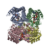 4ituC 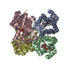 2cfcS S: Starting model for refinement C: citing same article ( |
|---|---|
| Similar structure data |


-Links
 PDBj
PDBj


- Assembly
| Deposited unit | 
| |||||||||||||||
|---|---|---|---|---|---|---|---|---|---|---|---|---|---|---|---|---|
| 1 |
| |||||||||||||||
| Unit cell |
| |||||||||||||||
| Components on special symmetry positions |
|
-Components
| #1: Protein | Mass: 27112.775 Da / Num. of mol.: 4 Source method: isolated from a genetically manipulated source Source: (gene. exp.) Xanthobacter autotrophicus (bacteria) / Strain: Py2 / Gene: Xaut_5073 / Plasmid: pET28-b / Production host: #2: Chemical | ChemComp-NAD /   Mass: 663.425 Da / Num. of mol.: 4 / Source method: obtained synthetically / Formula: C21H27N7O14P2 / Comment: NAD*YM Mass: 663.425 Da / Num. of mol.: 4 / Source method: obtained synthetically / Formula: C21H27N7O14P2 / Comment: NAD*YM#3: Chemical | ChemComp-ACT /   Mass: 59.044 Da / Num. of mol.: 4 / Source method: obtained synthetically / Formula: C2H3O2 Mass: 59.044 Da / Num. of mol.: 4 / Source method: obtained synthetically / Formula: C2H3O2#4: Water | ChemComp-HOH / |  Mass: 18.015 Da / Num. of mol.: 1027 / Source method: isolated from a natural source / Formula: H2O Mass: 18.015 Da / Num. of mol.: 1027 / Source method: isolated from a natural source / Formula: H2O |
|---|
-Experimental details
-Experiment
| Experiment | Method: X-RAY DIFFRACTION / Number of used crystals: 1 |
|---|
- Sample preparation
| Crystal | Density Matthews: 2 Å3/Da / Density % sol: 38.52 % |
|---|---|
| Crystal grow | Temperature: 277 K / Method: vapor diffusion, sitting drop / pH: 7.5 Details: 0.35M ammonium acetate, 27% polyethylene glycol, pH 7.5, VAPOR DIFFUSION, SITTING DROP, temperature 277K |
-Data collection
| Diffraction | Mean temperature: 80 K |
|---|---|
| Diffraction source | Source: ROTATING ANODE / Type: RIGAKU RU200 / Wavelength: 1.5418 Å |
| Detector | Type: RIGAKU RAXIS IV++ / Detector: IMAGE PLATE / Date: Sep 2, 2010 / Details: mirrors |
| Radiation | Monochromator: Ni filter / Protocol: SINGLE WAVELENGTH / Monochromatic (M) / Laue (L): M / Scattering type: x-ray |
| Radiation wavelength | Wavelength: 1.5418 Å / Relative weight: 1 |
| Reflection | Resolution: 1.6→35 Å / Num. all: 113574 / Num. obs: 113574 / % possible obs: 16 % / Observed criterion σ(F): 0 / Observed criterion σ(I): 0 / Redundancy: 6.2 % / Rmerge(I) obs: 0.063 / Rsym value: 0.046 / Net I/σ(I): 25.75 |
| Reflection shell | Resolution: 1.6→1.66 Å / Redundancy: 2.8 % / Rmerge(I) obs: 0.444 / Mean I/σ(I) obs: 1.92 / Num. unique all: 9812 / Rsym value: 0.423 / % possible all: 86 |
- Processing
| Software |
| |||||||||||||||||||||||||||||||||||||||||||||||||||||||||||||||||||||||||||||||||||||||||||||||||||||||||||||||||||||||||||||||||||||||||||||||||||||||||||||||||||||||||||||||||||||||||||||||||||||||||||||||||||||||||
|---|---|---|---|---|---|---|---|---|---|---|---|---|---|---|---|---|---|---|---|---|---|---|---|---|---|---|---|---|---|---|---|---|---|---|---|---|---|---|---|---|---|---|---|---|---|---|---|---|---|---|---|---|---|---|---|---|---|---|---|---|---|---|---|---|---|---|---|---|---|---|---|---|---|---|---|---|---|---|---|---|---|---|---|---|---|---|---|---|---|---|---|---|---|---|---|---|---|---|---|---|---|---|---|---|---|---|---|---|---|---|---|---|---|---|---|---|---|---|---|---|---|---|---|---|---|---|---|---|---|---|---|---|---|---|---|---|---|---|---|---|---|---|---|---|---|---|---|---|---|---|---|---|---|---|---|---|---|---|---|---|---|---|---|---|---|---|---|---|---|---|---|---|---|---|---|---|---|---|---|---|---|---|---|---|---|---|---|---|---|---|---|---|---|---|---|---|---|---|---|---|---|---|---|---|---|---|---|---|---|---|---|---|---|---|---|---|---|---|
| Refinement | Method to determine structure: MOLECULAR REPLACEMENT Starting model: PDB ENTRY 2CFC Resolution: 1.6→26.425 Å / SU ML: 0.23 / σ(F): 0 / Phase error: 18 / Stereochemistry target values: ML
| |||||||||||||||||||||||||||||||||||||||||||||||||||||||||||||||||||||||||||||||||||||||||||||||||||||||||||||||||||||||||||||||||||||||||||||||||||||||||||||||||||||||||||||||||||||||||||||||||||||||||||||||||||||||||
| Solvent computation | Shrinkage radii: 0.73 Å / VDW probe radii: 1 Å / Solvent model: FLAT BULK SOLVENT MODEL / Bsol: 32.254 Å2 / ksol: 0.358 e/Å3 | |||||||||||||||||||||||||||||||||||||||||||||||||||||||||||||||||||||||||||||||||||||||||||||||||||||||||||||||||||||||||||||||||||||||||||||||||||||||||||||||||||||||||||||||||||||||||||||||||||||||||||||||||||||||||
| Displacement parameters | Biso mean: 13.77 Å2
| |||||||||||||||||||||||||||||||||||||||||||||||||||||||||||||||||||||||||||||||||||||||||||||||||||||||||||||||||||||||||||||||||||||||||||||||||||||||||||||||||||||||||||||||||||||||||||||||||||||||||||||||||||||||||
| Refinement step | Cycle: LAST / Resolution: 1.6→26.425 Å
| |||||||||||||||||||||||||||||||||||||||||||||||||||||||||||||||||||||||||||||||||||||||||||||||||||||||||||||||||||||||||||||||||||||||||||||||||||||||||||||||||||||||||||||||||||||||||||||||||||||||||||||||||||||||||
| Refine LS restraints |
| |||||||||||||||||||||||||||||||||||||||||||||||||||||||||||||||||||||||||||||||||||||||||||||||||||||||||||||||||||||||||||||||||||||||||||||||||||||||||||||||||||||||||||||||||||||||||||||||||||||||||||||||||||||||||
| LS refinement shell |
|
