 Movie
Movie Controller

[English] 日本語
 Yorodumi
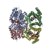
- PDB-2cfc: structural basis for stereo selectivity in the (R)- and (S)- hydr... -
+ Open data

- Basic information
| Entry | Database: PDB / ID: 2cfc | ||||||
|---|---|---|---|---|---|---|---|
| Title | structural basis for stereo selectivity in the (R)- and (S)- hydroxypropylethane thiosulfonate dehydrogenases | ||||||
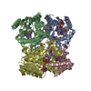
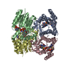
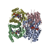
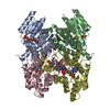
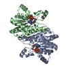
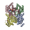
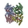
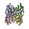
 Components Components | 2-(R)-HYDROXYPROPYL-COM DEHYDROGENASE | ||||||
 Keywords Keywords | OXIDOREDUCTASE / NAD | ||||||
| Function / homology |  Function and homology information Function and homology information2-(R)-hydroxypropyl-CoM dehydrogenase / 2-(R)-hydroxypropyl-CoM dehydrogenase activity / propylene catabolic process Similarity search - Function | ||||||
| Biological species |  XANTHOBACTER AUTOTROPHICUS (bacteria) XANTHOBACTER AUTOTROPHICUS (bacteria) | ||||||
| Method |  X-RAY DIFFRACTION / SYNCHROTRON / MAD / Resolution: 1.8 Å X-RAY DIFFRACTION / SYNCHROTRON / MAD / Resolution: 1.8 Å | ||||||
 Authors Authors | Krishnakumar, A.M. / Nocek, B.P. / Clark, D.D. / Ensign, S.A. / Peters, J.W. | ||||||
 Citation Citation | Journal: Biochemistry / Year: 2006 Title: Structural Basis for Stereoselectivity in the (R)-and (S)-Hydroxypropylthioethanesulfonate Dehydrogenases. Authors: Krishnakumar, A.M. / Nocek, B.P. / Clark, D.D. / Ensign, S.A. / Peters, J.W. | ||||||
| History |
|
- Structure visualization
| Structure viewer | Molecule: MolmilJmol/JSmol |
|---|
- Downloads & links
-Download
| PDBx/mmCIF format | 2cfc.cif.gz | 214.1 KB | Display | PDBx/mmCIF format |
|---|---|---|---|---|
| PDB format | pdb2cfc.ent.gz | 174 KB | Display | PDB format |
| PDBx/mmJSON format | 2cfc.json.gz | Tree view | PDBx/mmJSON format | |
| Others |  Other downloads Other downloads |
-Validation report
| Arichive directory | https://data.pdbj.org/pub/pdb/validation_reports/cf/2cfcftp://data.pdbj.org/pub/pdb/validation_reports/cf/2cfc | HTTPS FTP |
|---|
-Related structure data
| Similar structure data |
|---|

-Links
 PDBj
PDBj


- Assembly
| Deposited unit | 
| ||||||||
|---|---|---|---|---|---|---|---|---|---|
| 1 |
| ||||||||
| Unit cell |
| ||||||||
| Noncrystallographic symmetry (NCS) | NCS oper: (Code: given / Matrix: (1), |
-Components
| #1: Protein | Mass: 26165.854 Da / Num. of mol.: 4 / Source method: isolated from a natural source / Source: (natural) XANTHOBACTER AUTOTROPHICUS (bacteria) / Strain: PY2References: UniProt: Q56840, 2-(R)-hydroxypropyl-CoM dehydrogenase #2: Chemical | ChemComp-NAD /   Mass: 663.425 Da / Num. of mol.: 4 / Source method: obtained synthetically / Formula: C21H27N7O14P2 / Comment: NAD*YM Mass: 663.425 Da / Num. of mol.: 4 / Source method: obtained synthetically / Formula: C21H27N7O14P2 / Comment: NAD*YM#3: Chemical | ChemComp-KPC / (   Mass: 198.260 Da / Num. of mol.: 4 / Source method: obtained synthetically / Formula: C5H10O4S2 Mass: 198.260 Da / Num. of mol.: 4 / Source method: obtained synthetically / Formula: C5H10O4S2#4: Water | ChemComp-HOH / |  Mass: 18.015 Da / Num. of mol.: 915 / Source method: isolated from a natural source / Formula: H2O Mass: 18.015 Da / Num. of mol.: 915 / Source method: isolated from a natural source / Formula: H2O |
|---|
-Experimental details
-Experiment
| Experiment | Method: X-RAY DIFFRACTION / Number of used crystals: 1 |
|---|
- Sample preparation
| Crystal | Density Matthews: 2.34 Å3/Da / Density % sol: 47.36 % |
|---|---|
| Crystal grow | Details: 15MM NAD, 0.2M MAGNESIUM CHLORIDE, 0.1 M HEPES, PH7.5, 30% W/V PEG400 AND 0.1 SPERMINE TETRACHLORIDE AND 20MM R-HYDROXY PROPYL COM |
-Data collection
| Diffraction | Mean temperature: 77 K | |||||||||
|---|---|---|---|---|---|---|---|---|---|---|
| Diffraction source | Source: SYNCHROTRON / Site: SSRL  / Beamline: BL9-2 / Wavelength: 0.87008,1.00871 / Beamline: BL9-2 / Wavelength: 0.87008,1.00871 | |||||||||
| Detector | Type: MARRESEARCH / Detector: CCD / Details: MIRRORS | |||||||||
| Radiation | Monochromator: NI FILTER / Protocol: MAD / Monochromatic (M) / Laue (L): M / Scattering type: x-ray | |||||||||
| Radiation wavelength |
| |||||||||
| Reflection | Resolution: 1.8→20 Å / Num. obs: 149113 / % possible obs: 93.9 % / Observed criterion σ(I): 2 / Redundancy: 2.5 % / Rmerge(I) obs: 0.08 / Net I/σ(I): 6.5 | |||||||||
| Reflection shell | Highest resolution: 1.8 Å / Rmerge(I) obs: 0.3 / Mean I/σ(I) obs: 2.1 |
- Processing
| Software |
| ||||||||||||||||||||||||||||||||||||||||||||||||||||||||||||
|---|---|---|---|---|---|---|---|---|---|---|---|---|---|---|---|---|---|---|---|---|---|---|---|---|---|---|---|---|---|---|---|---|---|---|---|---|---|---|---|---|---|---|---|---|---|---|---|---|---|---|---|---|---|---|---|---|---|---|---|---|---|
| Refinement | Method to determine structure: MAD / Resolution: 1.8→50 Å / Data cutoff high absF: 10000 / Cross valid method: THROUGHOUT / σ(F): 0
| ||||||||||||||||||||||||||||||||||||||||||||||||||||||||||||
| Solvent computation | Bsol: 78.8068 Å2 / ksol: 0.384731 e/Å3 | ||||||||||||||||||||||||||||||||||||||||||||||||||||||||||||
| Displacement parameters |
| ||||||||||||||||||||||||||||||||||||||||||||||||||||||||||||
| Refinement step | Cycle: LAST / Resolution: 1.8→50 Å
| ||||||||||||||||||||||||||||||||||||||||||||||||||||||||||||
| Refine LS restraints |
| ||||||||||||||||||||||||||||||||||||||||||||||||||||||||||||
| Xplor file |
|
