 Movie
Movie Controller

[English] 日本語
 Yorodumi
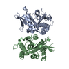
- PDB-4gc0: The structure of the MFS (major facilitator superfamily) proton:x... -
+ Open data

- Basic information
| Entry | Database: PDB / ID: 4gc0 | ||||||
|---|---|---|---|---|---|---|---|
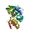
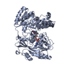
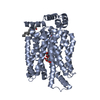
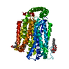
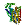
| Title | The structure of the MFS (major facilitator superfamily) proton:xylose symporter XylE bound to 6-bromo-6-deoxy-D-glucose | ||||||
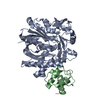
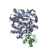
 Components Components | D-xylose-proton symporter | ||||||
 Keywords Keywords | TRANSPORT PROTEIN / MFS / D-xylose:proton symporter | ||||||
| Function / homology |  Function and homology information Function and homology informationD-xylose:proton symporter activity / D-xylose transmembrane transport / transmembrane transporter activity / transmembrane transport / membrane / plasma membrane Similarity search - Function | ||||||
| Biological species |  | ||||||
| Method |  X-RAY DIFFRACTION / SYNCHROTRON / MOLECULAR REPLACEMENT / Resolution: 2.6 Å X-RAY DIFFRACTION / SYNCHROTRON / MOLECULAR REPLACEMENT / Resolution: 2.6 Å | ||||||
 Authors Authors | Yan, N. / Sun, L.F. / Zeng, X. / Yan, C.Y. | ||||||
 Citation Citation | Journal: Nature / Year: 2012 Title: Crystal structure of a bacterial homologue of glucose transporters GLUT1-4. Authors: Sun, L. / Zeng, X. / Yan, C. / Sun, X. / Gong, X. / Rao, Y. / Yan, N. | ||||||
| History |
|
- Structure visualization
| Structure viewer | Molecule: MolmilJmol/JSmol |
|---|
- Downloads & links
-Download
| PDBx/mmCIF format | 4gc0.cif.gz | 200.6 KB | Display | PDBx/mmCIF format |
|---|---|---|---|---|
| PDB format | pdb4gc0.ent.gz | 161.1 KB | Display | PDB format |
| PDBx/mmJSON format | 4gc0.json.gz | Tree view | PDBx/mmJSON format | |
| Others |  Other downloads Other downloads |
-Validation report
| Arichive directory | https://data.pdbj.org/pub/pdb/validation_reports/gc/4gc0ftp://data.pdbj.org/pub/pdb/validation_reports/gc/4gc0 | HTTPS FTP |
|---|
-Related structure data
| Related structure data |  4gbySC  4gbzC S: Starting model for refinement C: citing same article ( |
|---|---|
| Similar structure data |

-Links
 PDBj
PDBj


- Assembly
| Deposited unit | 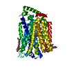
| ||||||||
|---|---|---|---|---|---|---|---|---|---|
| 1 |
| ||||||||
| Unit cell |
|
-Components
| #1: Protein | Mass: 53642.629 Da / Num. of mol.: 1 Source method: isolated from a genetically manipulated source Source: (gene. exp.) | ||
|---|---|---|---|
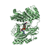
| #2: Sugar | ChemComp-6BG /   Type: D-saccharide, beta linking / Mass: 243.053 Da / Num. of mol.: 1 / Source method: obtained synthetically / Formula: C6H11BrO5 Type: D-saccharide, beta linking / Mass: 243.053 Da / Num. of mol.: 1 / Source method: obtained synthetically / Formula: C6H11BrO5 | ||
| #3: Sugar | ChemComp-BNG / 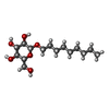  Type: D-saccharide / Mass: 306.395 Da / Num. of mol.: 4 Type: D-saccharide / Mass: 306.395 Da / Num. of mol.: 4Source method: isolated from a genetically manipulated source Formula: C15H30O6 / Comment: detergent*YM #4: Water | ChemComp-HOH / |  Mass: 18.015 Da / Num. of mol.: 7 / Source method: isolated from a natural source / Formula: H2O Mass: 18.015 Da / Num. of mol.: 7 / Source method: isolated from a natural source / Formula: H2O |
-Experimental details
-Experiment
| Experiment | Method: X-RAY DIFFRACTION / Number of used crystals: 1 |
|---|
- Sample preparation
| Crystal | Density Matthews: 3.62 Å3/Da / Density % sol: 66.05 % |
|---|---|
| Crystal grow | Temperature: 291 K / Method: vapor diffusion, hanging drop / pH: 9.6 Details: 40 % (w/v) PEG400, 0.05 M Glycine pH 9.6 and 0.1 M LiCl , VAPOR DIFFUSION, HANGING DROP, temperature 291K |
-Data collection
| Diffraction | Mean temperature: 100 K |
|---|---|
| Diffraction source | Source: SYNCHROTRON / Site: SSRF  / Beamline: BL17U / Wavelength: 0.9196 Å / Beamline: BL17U / Wavelength: 0.9196 Å |
| Detector | Type: ADSC QUANTUM 315r / Detector: CCD / Date: Mar 5, 2012 |
| Radiation | Monochromator: Si 111 CHANNEL / Protocol: SINGLE WAVELENGTH / Monochromatic (M) / Laue (L): M / Scattering type: x-ray |
| Radiation wavelength | Wavelength: 0.9196 Å / Relative weight: 1 |
| Reflection | Resolution: 2.6→40 Å / Num. all: 25081 / Num. obs: 24981 / % possible obs: 99.6 % / Observed criterion σ(F): 1 / Observed criterion σ(I): 1 |
| Reflection shell | Resolution: 2.6→2.69 Å / % possible all: 97.9 |
- Processing
| Software |
| |||||||||||||||||||||||||||||||||||||||||||||||||||||||||||||||||||||||||||||||||||||||||||||||||||||||||||||||||||||||
|---|---|---|---|---|---|---|---|---|---|---|---|---|---|---|---|---|---|---|---|---|---|---|---|---|---|---|---|---|---|---|---|---|---|---|---|---|---|---|---|---|---|---|---|---|---|---|---|---|---|---|---|---|---|---|---|---|---|---|---|---|---|---|---|---|---|---|---|---|---|---|---|---|---|---|---|---|---|---|---|---|---|---|---|---|---|---|---|---|---|---|---|---|---|---|---|---|---|---|---|---|---|---|---|---|---|---|---|---|---|---|---|---|---|---|---|---|---|---|---|---|
| Refinement | Method to determine structure: MOLECULAR REPLACEMENT Starting model: 4GBY Resolution: 2.6→39.123 Å / SU ML: 0.28 / σ(F): 1.12 / Phase error: 28.1 / Stereochemistry target values: ML
| |||||||||||||||||||||||||||||||||||||||||||||||||||||||||||||||||||||||||||||||||||||||||||||||||||||||||||||||||||||||
| Solvent computation | Shrinkage radii: 0.86 Å / VDW probe radii: 1.1 Å / Solvent model: FLAT BULK SOLVENT MODEL / Bsol: 67.919 Å2 / ksol: 0.343 e/Å3 | |||||||||||||||||||||||||||||||||||||||||||||||||||||||||||||||||||||||||||||||||||||||||||||||||||||||||||||||||||||||
| Displacement parameters |
| |||||||||||||||||||||||||||||||||||||||||||||||||||||||||||||||||||||||||||||||||||||||||||||||||||||||||||||||||||||||
| Refinement step | Cycle: LAST / Resolution: 2.6→39.123 Å
| |||||||||||||||||||||||||||||||||||||||||||||||||||||||||||||||||||||||||||||||||||||||||||||||||||||||||||||||||||||||
| Refine LS restraints |
| |||||||||||||||||||||||||||||||||||||||||||||||||||||||||||||||||||||||||||||||||||||||||||||||||||||||||||||||||||||||
| LS refinement shell |
| |||||||||||||||||||||||||||||||||||||||||||||||||||||||||||||||||||||||||||||||||||||||||||||||||||||||||||||||||||||||
| Refinement TLS params. | Method: refined / Origin x: -27.2331 Å / Origin y: 21.9288 Å / Origin z: -3.9812 Å
| |||||||||||||||||||||||||||||||||||||||||||||||||||||||||||||||||||||||||||||||||||||||||||||||||||||||||||||||||||||||
| Refinement TLS group | Selection details: all |
